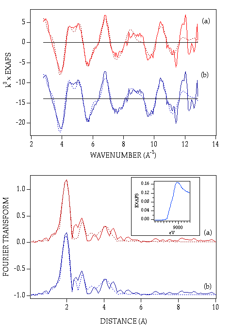
Figure 9. Experimental vs simulated EXAFS and Fourier
transforms of oxidized
DProPHM-382s. Upper panel: background-subtracted EXAFS. Lower panel:
Fourier
transforms. (a) Spectrum simulated with 2.5 His, 1.5 O/N scatterers
with
parameters as given in Table 2. (b) Spectrum simulated as in (a) with
inclusion
of a shell of 0.3 S at 2.40 Angstrom.
Back
to PHM paper