5. Electron Paramagnetic Resonance Study of the Reaction of Chromium(V) Species with Reducing Agents
Since the studies discussed in previous chapters showed that relatively stable chromium(V) complexes were formed during the reduction of chromium(VI) by a number of thiols, further experiments were performed in an attempt to characterize possible chromium(V)-thiol complexes, and to study the reactivity of chromium(V) towards thiols. Toward this end, relatively stable chromium(V) complexes such as bis(2-hydroxy-2-methylbutyrato)oxochromate(V) and bis(2-ethyl-2-hydroxybutyrato)oxochromate(V) were used as the initial form of chromium in further reactions involving thiols. Substitution of thiol for one or more butyrato ligands on the chromium(V) center and reduction of chromium(V) by thiol were proposed as a possibilities in these reactions.
5.1. Results and Discussion
5.1.1.
b-mercaptoethanol
The reaction of sodium bis(2-hydroxy-2-methylbutyrato)oxochromate(V) ((HMBA)
2Cr(V)) (50.0 mM) and b-mercaptoethanol (50.0 mM) in aqueous solution at an initial pH = 5.9 (not adjusted during the reaction) and 25 °C was followed via frozen solution (77K) EPR spectroscopy. Figure 5-1 shows the spectra obtained during this reaction. It should be noted that the starting chromium(V) complex, (HMBA)2Cr(V), EPR spectrum is much more intense than subsequent EPR spectra. Within 1.2 minutes, 98.6% of the initial (HMBA)2Cr(V) has been reduced to chromium(III). Figure 5-1-A shows the small amount of chromium(V) species left after 1.2 minutes, with a single signal at g = 1.982 (DH = 68 G). This g-value and linewidth is identical to that of the starting chromium(V) complex. It is interesting to note that under the conditions employed the spectra due to chromium(III) was clearly evident in the g = 2.5-5.0 region (Figure 5-1)-B ((HMBA)2Cr(V) spectrum is not shown for clarity; no Cr(III) features evident).
These data show that reduction of the starting chromium(V) complex is much more favorable than ligand substitution under these conditions. Most of the chromium(V) (98.6%) is reduced to chromium(III) within the first minute of reaction. Once the chromium is reduced, some slow ligand substitution takes place, as evidenced by the change in the chromium(III) signals in the g = 2.5-5 region (Figure 5-1). Stock bis(2-hydroxy-2-methylbutyrato)oxochromate(V) in the absence of
b-mercaptoethanol shows no evidence of chromium(III) in the same region of the spectrum over the same time period as the reaction with b-mercaptoethanol.
The reaction of sodium bis(2-hydroxy-2-methylbutyrato)oxochromate(V) (5.0 mM) and
b-mercaptoethanol (5.0 mM) in 1:1 acetic acid:water solution and -15 °C was followed via frozen solution (77K) and solution EPR spectroscopy. Figure 5-2-A shows the frozen solution EPR spectra of the reaction, with a single chromium(V) species evident at g = 1.980 (DH = 24 G). Again, the chromium(V) signals at later time points are identical to the starting chromium(V) complex. Under the conditions employed, the reduction of the chromium(V) complex is slower than in aqueous solution at pH 5.9 (above). A very small amount of chromium(III) is observed in the EPR spectra under these conditions (Figure 5-2)-B.
Figure 5-3 shows the solution EPR spectra obtained for the reaction of sodium bis(2-hydroxy-2-methylbutyrato)oxochromate(V) (5.0 mM) and
b-mercaptoethanol (5.0 mM) in 1:1 acetic acid:water solution and -15 °C. There is a slight shift in g-value from 1.9798 (DH = 1.17 G) to 1.9802 (DH = 1.27 G) in the first minute of the reaction, and then only reduction of the chromium(V) center subsequently occurs. Also, there is a slight shift in the observed hyperfine values with a53Cr = 18.48 G for the starting chromium(V) complex, and a53Cr = 18.18 G for the reaction product. These data are consistent with simple reduction of the chromium(V) center. It is possible that ligand substitution occurs immediately upon introduction of the thiol, and then subsequent reduction of the chromium(V) center occurs. While the increase in g-value and decrease in hyperfine are consistent with the introduction of sulfur into the coordination sphere of the chromium(V) center, the absolute changes in these values are not large enough to decisively conclude that ligand substitution has occurred.
Figure 5-4 shows the solution EPR spectra obtained for the reaction of sodium bis(2-hydroxy-2-methylbutyrato)oxochromate(V) (5.0 mM) and b-mercaptoethanol (5.0 mM) in 1:1 acetic acid:water solution and -15 °C. This is the same reaction as outlined above, however the EPR spectra were obtained with a smaller (0.1 G vs. 1 G) modulation amplitude to detect any superhyperfine that may be observable due to the presence of b-mercaptoethanol. The spectra again show an initial slight change in g-value from 1.9798 (DH = 1.08 G) to 1.9799 (DH = 1.08 G), and a slight change in hyperfine from a53Cr = 18.57 G to 18.38 G. The only new feature presented was an increase in the resolution of the splitting of the outer a53Cr hyperfine satellites.
Figure 5-5 shows the solution EPR spectra obtained for the reaction of sodium bis(2-hydroxy-2-methylbutyrato)oxochromate(V) (5.0 mM) and b-mercaptoethanol (5.0 mM) in 1:1 acetic acid:water solution with an ionic strength of 1.5 (maintained via KCl) and -15 °C. The spectra again show an initial slight change in g-value from 1.9800 (DH = 0.98 G) to 1.9801 (DH = 0.98 G), and no change in hyperfine of 18.47 G. The increased ionic strength appears to have slowed the reduction of the chromium(V) center, since only about 50% reduction has occurred in about 8 minutes at I = 1.5 (Figure 5-5) while at lower ionic strength the reduction was about 80% completed in about 6.5 minutes (Figure 5-4).
Higher thiol:chromium ratios were deemed necessary to effect ligand substitution in this reaction, so a 20:1 thiol:chromium ratio was attempted. The reaction of sodium bis(2-hydroxy-2-methylbutyrato)oxochromate(V) (8.0 mM) and b-mercaptoethanol (160.0 mM) in 1:1 acetic acid:water solution at -15 °C resulted in complete reduction of chromium(V) to chromium(III) upon mixing; no chromium(V) was observed in as little as 45 seconds reaction time. The reaction of sodium bis(2-hydroxy-2-methylbutyrato)oxochromate(V) (5.0 mM) and b-mercaptoethanol (10.0 mM; 1:2 chromium:thiol ratio) in 1:1 acetic acid:water solution at -15 °C resulted in 91.7% reduction of the starting chromium(V) complex within 1.3 minutes (Figure 5-6). Under the condition employed it is apparent that reduction of the chromium(V) center has occurred without ligand substitution since the g-value of the reaction products, g = 1.9799 (DH = 1.08 G) is not significantly different from the starting chromium(V) complex with g = 1.9797 (DH = 1.17 G) (Figure 5-6).
Since ligand substitution of b-mercaptoethanol onto the chromium(V) center of bis(2-hydroxy-2-methylbutyrato)oxochromate(V) was not observed in aqueous solutions, non-aqueous reaction conditions were employed. It has been suggested that reduction of bis(2-hydroxy-2-methylbutyrato)oxochromate(V) may be preceded by hydrolysis (46), thus a dry acetone solvent would most likely retard the reduction of the complex.
The reaction of sodium bis(2-hydroxy-2-methylbutyrato)oxochromate(V) (5.0 mM) and b-mercaptoethanol (10.0 mM; 1:2 ratio of chromium:thiol) in acetone at 25 °C was followed via solution EPR spectroscopy. Figure 5-7 shows the EPR spectra obtained. The starting complex and the subsequent spectra have the same g-value of 1.9799. The spectra show very little change over time; little or no reduction of the chromium(V) center occurs over a 20 minute reaction time. An interesting result is that at about 20 minutes into the reaction, the central chromium(V) feature has the same g-value of 1.9799, but the lineshape has become noticeably sharper. Also, the satellite a53Cr hyperfine feature at higher field shows a dramatic change in lineshape, with a splitting of the signal becoming very apparent (Figure 5-7).
The reaction of sodium bis(2-hydroxy-2-methylbutyrato)oxochromate(V) (5.0 mM) and b-mercaptoethanol (100.0 mM; 1:20 chromium:thiol ratio) in acetone at 25 °C was followed via solution EPR spectroscopy. Figure 5-8 shows the EPR spectra obtained. The starting complex and the subsequent spectra have the same g-value of 1.9799. Within 4 minutes of the start of the reaction a significant change in the linewidth of the chromium(V) signal occurs, changing from 1.955 G for the starting chromium(V) compound to 1.564 G for the reaction species. Reduction of the chromium(V) species occurs slowly, with only about 55% reduction in about 20 minutes. Significant chromium(V) is observed more than 60 minutes after the start of the reaction. As the reaction proceeds, the a53Cr hyperfine satellite signal becomes more significantly split (Figure 5-8).
The reaction of sodium bis(2-hydroxy-2-methylbutyrato)oxochromate(V) (5.0 mM) and b-mercaptoethanol (500.0 mM; 1:100 chromium:thiol ratio) in acetone at 25 °C was followed via solution EPR spectroscopy. Figure 5-9 shows the EPR spectra obtained. The starting complex and the subsequent spectra have the same g-value of 1.9799. Within 1 minute of the start of the reaction a significant change in the linewidth of the chromium(V) signal occurs, changing from 1.955 G for the starting chromium(V) compound to 1.467 G for the reaction species. Reduction of the chromium(V) species occurs relatively quickly, with about 90% reduction in about 15.5 minutes. As the reaction proceeds, the a53Cr hyperfine satellite signal becomes more significantly split (Figure 5-9-B).
5.1.1.1. Discussion
It seems clear from these data that in aqueous (either H
2O or 50% acetic acid) solutions the reaction of sodium bis(2-hydroxy-2-methylbutyrato)oxochromate(V) and b-mercaptoethanol does not result in any significant ligand substitution reaction, but rather simple reduction of the parent chromium(V) complex by the thiol. The main EPR signal remains relatively consistent in lineshape and g-value (~1.980). The g- values observed upon addition of thiol do not increase as would be expected for a change in coordination from O to S on the chromium center. In a non-aqueous solvent such as acetone however, the reduction of the parent chromium(V) complex is greatly retarded, even at high thiol:chromium ratios (100:1), and a number of interesting spectral features develop in the EPR spectra. Most notable is the ~20% smaller linewidth of the central
chromium(V) signal upon addition of thiol, while the g-value remains constant at g = 1.9799. Also of note is the significant splitting of the a
53Cr hyperfine satellites at higher field for the reactions done in acetone. Bramley et al. (72) have reported a solvent dependence of the EPR spectra of oxochromate(V) complexes, and have attributed the splitting of the 53Cr satellites in weakly hydrogen-bonding solvents such as acetone to protonation of the alcohol group of the ehba ligand. Splitting of the central 53Cr satellites is not observed, and is most likely due to a fast-exchange coalescence involving the protonation/deprotonation equilibrium (72).
5.1.2. Dithiothreitol
The reaction of potassium perchromate, K
3CrO8 (5.0 mM) and dithiothreitol (15.0 mM) in 0.2 N KOH (aqueous) at 25 °C was followed via solution EPR spectroscopy. Upon addition of thiol, no significant changes in the EPR spectra (g = 1.974; DH = 1.76 G; a53Cr = 19.9 G) occur, and the EPR signal remains constant for over an hour, with subsequent slow loss of chromium(V) signal over 24 hours (data shown only up to 13 minutes) Figure 5-10. This loss of chromium(V) signal follows the same time-course as K3CrO8 in 0.2 N KOH (stock), i.e., the presence of dithiothreitol appears to have no effect on the perchromate species under these conditions.
The reaction of sodium bis(2-ethyl-2-hydroxybutyrato)oxochromate(V) (5.0 mM) and dithiothreitol (10.0 mM) in 1:1 Acetic acid:water solution at 25 °C was followed via solution EPR spectroscopy. The solution EPR spectra of the reaction shows the initial chromium(V) complex (g = 1.980;
DH = 0.98 G; a53Cr = 18.3 G) (Figure 5-11). Within 5 minutes following addition of dithiothreitol, about 95% of the chromium(V) signal is lost; it is clear from these data that under these conditions the reduction of the starting chromium(V) species occurs very quickly, and no ligand substitution takes place. Increasing the dithiothreitol concentration to 100.0 mM (1:20 Cr:dithiothreitol ratio) results in complete reduction of the starting chromium(V) species upon mixing.
The reaction of sodium bis(2-hydroxy-2-methylbutyrato)oxochromate(V) (5.0 mM) and dithiothreitol (100.0 mM; 1:20 Cr:dithiothreitol ratio) in acetone at 25 °C was followed via solution EPR spectroscopy. Figure 5-12 shows the solution EPR spectra of the reaction, with two signals: g = 1.980 (due to the starting chromium(V) species (main signal)) and g(peak) = 1.983 (due to a new chromium(V) species) evident. The a53Cr hyperfine value for the g = 1.980 signal is 17.89 G for the starting chromium(V) complex and for the reaction. Upon addition of thiol, the linewidth of the g = 1.980 signal changes, narrowing from 1.76 G to 1.43 G. Also, upon addition of thiol, the a53Cr hyperfine satellite at higher field is significantly split (Figure 5-12-B). The most interesting observation is the formation of a new signal with a peak at g(peak) = 1.983. This new signal is not totally resolved due to the intensity of the g = 1.980 signal, however it is clear that a new chromium(V) species is formed, presumably by substitution of dithiothreitol for one of the butyrato ligands. Both signals show a decay over time, with about 93% loss of chromium(V) signals within 10 minutes.
The reaction of sodium bis(2-ethyl-2-hydroxybutyrato)oxochromate(V) (5.0 mM) and dithiothreitol (100.0 mM; 1:20 Cr:dithiothreitol ratio) in acetonitrile at 25 °C was followed via solution EPR spectroscopy. Figure 5-13 shows the solution EPR spectra of the initial chromium(V) complex and the reaction with dithiothreitol; one main signal at g = 1.980 is observed. The a53Cr hyperfine value for the g = 1.980 signal is 17.89 G for the starting chromium(V) complex and for the reaction (Figure 5-13-C). Upon addition of thiol, the linewidth of the g = 1.980 signal changes, narrowing from 1.66 G to 1.46 G in 3 minutes, and narrowing even more to 1.07 G at 22 minutes. Also, upon addition of thiol, the a53Cr hyperfine satellite at higher field is significantly split, while the starting chromium(V) species alone is not split in this solvent (Figure 5-13-C). The chromium(V) signal decays relatively slowly over time, with about 98% loss of chromium(V) signals within 20 minutes (Figure 5-13-B).
The reaction of sodium bis(2-ethyl-2-hydroxybutyrato)oxochromate(V) (5.0 mM) and dithiothreitol (25.0 mM; 1:5 Cr:dithiothreitol ratio) in N,N-dimethylformamide at 25 °C was followed via solution EPR spectroscopy. Figure 5-14 shows the solution EPR spectra of the reaction, with one main signal at g = 1.980 corresponding to the starting chromium(V) complex and the result of addition of dithiothreitol. The a53Cr hyperfine value for the g = 1.980 signal is 17.8 G for the starting chromium(V) complex and for the reaction (Figure 5-14-B). A new chromium(V) signal at gpeak = 1.983 is observed after 40 minutes. Upon addition of thiol, the linewidth of the g = 1.980 signal changes, narrowing from 1.17 G to 1.08 G in 1 minute, and narrowing even more to 0.88 G at 41 minutes. Unlike the other solvents studied, upon addition of thiol the a53Cr hyperfine satellite at higher field is not split (Figure 5-14-B). The chromium(V) signal is quite stable, with about 98% retention of integrated chromium(V) signal after 40 minutes.
The reaction of sodium bis(2-ethyl-2-hydroxybutyrato)oxochromate(V) (5.0 mM) and dithiothreitol (7.5, 25.0 or 100.0 mM; 1:1.5, 5, or 20 Cr:dithiothreitol ratio) in methanol at 25 °C was followed via solution EPR spectroscopy. Figure 5-15 shows the solution EPR spectra of the 1:20 (chromium:dithiothreitol) reaction, with signals at g = 1.980 (due to the starting chromium(V) species (main signal)), and g(peak) = 1.984, 1.988 (due to two new chromium(V) species) evident. The a53Cr hyperfine value for the g = 1.980 signal is 18.2 G for the starting chromium(V) complex and for the reaction. Upon addition of thiol, the linewidth of the g = 1.980 signal changes, narrowing from 1.46 G to 1.37 G within 1 minute, and narrowing further to 0.88 G in 6 minutes. Also, upon addition of thiol, the a53Cr hyperfine satellite at higher field is significantly split (Figure 5-15-B). The most interesting observation is the formation of two new signals with g(peak) = 1.984 and g = 1.988. The new signals are not totally resolved due to the intensity of the g = 1.980 signal, however it is clear that new chromium(V) species are formed, presumably by substitution of dithiothreitol for one or both of the butyrato ligands. All chromium(V) signals show a decay over time, with total loss of chromium(V) signals within 10 minutes. The 1:5 (chromium:dithiothreitol) (Figure 5-16) or 1:1.5 (Figure 5-17) reactions result in the same EPR signals as the 1:20 reaction, however the decay of the chromium(V) signal is slower; 99% loss of signal at 40 minutes for 1:5 and 48% loss of signal at 125 minutes for the 1:1.5 ratio.
Figure 5-18 shows the time-course for the main g = 1.980 EPR signal observed in the reaction of sodium bis(2-ethyl-2-hydroxybutyrato)oxochromate(V) (5.0 mM) and dithiothreitol in methanol at the three Cr:dithiothreitol ratios studied (1:20, 5, 1.5). It is clear from these data that the rate of loss of chromium(V) signal is dependent on the dithiothreitol concentration; the higher thiol concentration effects the fastest rate. Also, these data seem to indicate that the chromium(V) signal observed is due to changing chromium(V) species, as evidenced by the oscillatory behavior observed at earlier time points in these reactions (Figure 5-18, inset). Further evidence for changing chromium(V) species is shown in Figure 5-17-B where the feature at g = 1.977 is initially present, blends into the main g = 1.980 signal at 5.5 minutes, and then re-appears later in the reaction. Loss and subsequent reappearance of this feature clearly indicates a change in the ligand environment around the chromium(V) center. Also, the loss of the g = 1.977 feature coincides with the appearance of the g(peak) = 1.984 species.
The feature at g = 1.977 is the result of a solvent effect on the bis(2-ethyl-2-hydroxybutyrato)oxochromate(V) species (as it is present in the absence of added thiol). Figure 5-19 shows the effect of solvent on the solution EPR spectrum of sodium bis(2-ethyl-2-hydroxybutyrato)oxochromate(V). These spectra show that the chromium(V) signal observed in acetic acid and methanol solvents displays a splitting of the a53Cr hyperfine satellite at high field, whereas dimethylformamide and acetonitrile do not show a splitting of the satellite. It should be noted that the chromium(V) signal in methanol effects the most extensive splitting of the satellite, and is the only solvent to display a feature at g = 1.977 (Figure 5-19).
Since the reactions of sodium bis(2-ethyl-2-hydroxybutyrato)oxochromate(V) with dithiothreitol in methanol yielded two new chromium(V) signals, a comparison reaction using chromium(VI) and dithiothreitol under the same conditions was performed. Figure 5-20 shows the solution EPR spectra resulting from the reaction of sodium dichromate (5.0 mM Cr(VI)) and dithiothreitol (25.0 mM; 1:5 Cr:dithiothreitol ratio) in methanol at 25 °C. At about 6 minutes into the reaction, four signals are observed, at g = 1.982, 1.984, 1.988, and g(peak) = 1.986 (Figure 5-20-A). At about 14 minutes into the reaction, the intermediate g(peak) = 1.986 signal is no longer present, and only three signals are observed, at g = 1.982, 1.984, and 1.988, (Figure 5-20-B). It is clear from these data that the g = 1.988 and g(peak) = 1.984 species observed upon reaction of bis(2-ethyl-2-hydroxybutyrato)oxochromate(V) with dithiothreitol (Figures 3-15, 16, 17) arise from a chromium(V)-dithiothreitol complex, as they are also observed in the reaction involving chromium(VI). The g = 1.982 feature observed in the chromium(VI) reaction is not observed in the chromium(V) reactions; it may be masked by the intense g = 1.980 signal due to bis(2-ethyl-2-hydroxybutyrato)oxochromate(V).
5.1.2.1. Discussion
Comparison of the EPR spectra of bis(2-ethyl-2-hydroxybutyrato)oxochromate(V) in various solvents (Figure 5-19) shows that the more strongly acidic solvents (acetic acid and methanol) result in splitting of the
53Cr satellites at higher field. This has been reported by Bramley et al. (72) to be due to protonation of the 2-ethyl-2-hydroxybutyrato ligand via solvent, and this work shows that the protonation of the ligand does not occur in N,N-dimethylformamide, and this is consistent with the need for a protic solvent. Also, the new feature at g = 1.977 (Figure 5-19) when methanol is used as a solvent has been assigned to (2-ethyl-2-hydroxybutanoato(2-))tris(methanolato(1-)oxochromate(V) (72).
The reaction of bis(2-ethyl-2-hydroxybutyrato)oxochromate(V) and dithiothreitol (1:2 ratio) in acetic acid solvent merely resulted in fast reduction of the starting chromium(V) complex, and no indication of new chromium(V) species was observed. The same reaction performed in acetonitrile also resulted in only the reduction of the starting chromium(V) complex. In contrast, the reaction of bis(2-ethyl-2-hydroxybutyrato)oxochromate(V) and dithiothreitol in acetone (1:20 ratio) or N,N-dimethylformamide (1:5 ratio) resulted in a much slower reduction of the starting chromium(V) complex, and the formation of a new chromium(V) signal at g
peak = 1.983 within 1.2 minutes of addition of thiol. Also, upon addition of thiol, the 53Cr satellites at higher field were significantly split. In methanol (1:20 ratio), the reduction of the starting chromium(V) complex was complete within 10 minutes, two new chromium(V) signals (g = 1.984, 1.988) were observed 1.25 minutes after addition of thiol and persisted until most of the initial chromium(V) (g = 1.980) was reduced. At a lower chromium(V):dithiol ratio (1:5), the two new signals at g = 1.984 and g = 1.988 were more intense, and reached a maximum 9 minutes after addition of thiol. These chromium(V) signals were observed for up to 2 hours when the chromium(V):dithiol ratio was 1:1 (less than stoichiometric amount for complete reduction of all starting chromium(V)). The new chromium(V) signals are most likely due to ligand substitution on the chromium(V) center, with one or both butyrato ligands being replaced by dithiothreitol. Upon addition of thiol, one of the butyrato ligands on bis(2-ethyl-2-hydroxybutyrato)oxochromate(V) (XV) is replaced by dithiothreitol, resulting in (XVI), with an EPR signal at g = 1.984. In methanol, (XVI) persists long enough for another dithiothreitol moiety to replace the remaining butyrato ligand, resulting in a bis-(dithiothreitol)chromium(V) complex (XVII) g = 1.988:
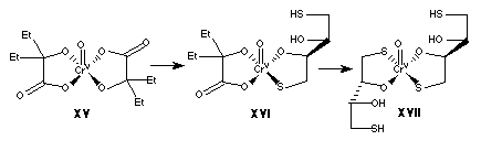 . Convincing evidence that the new features observed in the reaction of bis(2-ethyl-2-hydroxybutyrato)oxochromate(V) with dithiothreitol in methanol are due to mono- and bis-(dithiothreitol)chromium(V) complexes is found in the EPR spectra for the reaction of sodium dichromate (chromium(VI)) with dithiothreitol in methanol under the same conditions. Three features in the EPR spectra are observed for up to 13 minutes after addition of thiol, and two of these features (g = 1.984, 1.988) are at the same g-values as the new features observed in the reactions starting with chromium(V). Since no 2-ethyl-2-hydroxybutyrato ligands are present in reactions involving chromium(VI), the features at g = 1.984, 1.988 are assigned to (
XVI) and (XVII), respectively.
5.1.3. Glutathione
The reaction of sodium bis(2-hydroxy-2-methylbutyrato)oxochromate(V) (5.0 mM) and glutathione (5.0 mM; 1:1 Cr:glutathione ratio) in 1:1 acetic acid:water solution at 25 °C was followed via solution EPR spectroscopy. The initial bis(2-hydroxy-2-methylbutyrato)oxochromate(V) species showed a single chromium(V) signal at g = 1.980 (
DH = 1.08 G; a53Cr = 18.53 G) (Figure 5-21). Upon addition of thiol a slow loss of the chromium(V) signal was observed. It is clear from these data that under these conditions the reduction of the starting chromium(V) species occurs without ligand substitution, as the chromium(V) signal observed in the presence of thiol is identical to the starting chromium(V) complex, but less intense. Decreasing the sodium bis(2-hydroxy-2-methylbutyrato)oxochromate(V) concentration to 0.5 mM (1:10 Cr:glutathione ratio) also resulted in reduction of the starting chromium(V) species over time, however a new, weak signal at gpeak = 1.983 was observed 1.2 minutes after addition of thiol (Figure 5-22). When the Cr:glutathione ratio was changed to 1:3 (5.0 mM sodium bis(2-hydroxy-2-methylbutyrato)oxochromate(V): 15 mM glutathione), and the reaction is performed at -15 °C, the gpeak = 1.983 signal was not present, and only the slow loss of chromium(V) signal was observed (Figure 5-23).
The reaction of sodium bis(2-ethyl-2-hydroxybutyrato)oxochromate(V) (5.0 mM) and glutathione (100.0 mM; 1:20 Cr:glutathione ratio) in acetone at 25 °C was followed via solution EPR spectroscopy. The initial bis(2-ethyl-2-hydroxybutyrato)oxochromate(V) species showed a single chromium(V) signal at g = 1.980 (
DH = 1.66 G; a53Cr = 17.79 G) (Figure 5-24). Upon addition of thiol an initial decrease in intensity was observed for the chromium(V) signal while the g, linewidth, and hyperfine values did not change significantly; however, further loss of signal was not observed over 70 minutes (Figure 5-24). It should be noted that glutathione is only sparingly soluble in acetone, so this reaction was done as an inhomogeneous slurry. It is clear from these data that neither ligand substitution nor significant reduction of the starting chromium(V) species occurred under the conditions employed.
The reaction of sodium bis(2-ethyl-2-hydroxybutyrato)oxochromate(V) (5.0 mM) and glutathione (100.0 mM; 1:20 Cr:glutathione ratio) in N,N-dimethylformamide at 25 °C was followed via solution EPR spectroscopy. The initial bis(2-ethyl-2-hydroxybutyrato)oxochromate(V) species showed a single chromium(V) signal at g = 1.980 (DH = 1.17 G; a53Cr = 17.79 G) (Figure 5-25). Upon addition of thiol, no significant change in the EPR spectrum was observed over 120 minutes (Figure 5-25). It should be noted that glutathione is only sparingly soluble in N,N-dimethylformamide, so this reaction was done as an inhomogeneous slurry. It is clear from these data that neither ligand substitution nor significant reduction of the starting chromium(V) species occurred under the conditions employed.
5.1.3.1. Discussion
It is clear from these data that in non-aqueous solvents the reaction of bis(2-ethyl-2-hydroxybutyrato)oxochromate(V) with glutathione results in an initial slight loss of chromium(V) signal intensity, and then the EPR spectra are constant for up to 2 hours. Since glutathione is sparingly soluble in non-aqueous solvents, the chromium(V):glutathione ratio in these reactions was less than 1:20, nevertheless, sufficient glutathione would be available to reduce chromium(V) to chromium(III), as the reactions were performed as an inhomogenous slurry.
In contrast, the reaction of bis(2-ethyl-2-hydroxybutyrato)oxochromate(V) with glutathione in 50% acetic acid did result in a slow reduction of the starting chromium(V) complex at 1:1 and 1:3 chromium:glutathione ratios, with no new chromium(V) signals observed. At a low chromium(V) concentration, but at a 1:10 ratio, the rate of reduction of the initial chromium(V) complex was accelerated, and a new, very low-intensity signal was observed at g = 1.983. This new signal may be due to exchange of one of the butyrato ligands for glutathione, resulting in a (2-ethyl-2-hydroxybutyrato)(glutathione)oxochromium(V) complex.
5.1.4. Oxidized Glutathione
The reaction of sodium bis(2-hydroxy-2-methylbutyrato)oxochromate(V) (5.0 mM) and oxidized glutathione (GSSG) (50.0 mM; 10:1 Cr:GSSG ratio) in aqueous solution at 25 °C (initial pH = 3.8) was followed via solution EPR spectroscopy. The initial bis(2-hydroxy-2-methylbutyrato)oxochromate(V) species showed a single chromium(V) signal at g = 1.980 (
DH = 1.17 G; a53Cr = 18.57 G) (Figure 5-26). Upon addition of GSSG a slow loss of the chromium(V) signal was observed, and the linewidth of the chromium(V) signal increased to 1.37 G. When the reaction was followed via frozen solution (77 K) EPR, a single isotropic chromium(V) signal was observed at g = 1.977 for both the reaction spectra and the starting chromium(V) complex, and this signal slowly decreased with time (Figure 5-27). The signal at g = 5.24 signal observed in the 77K EPR spectra is not present in the starting chromium(V) complex; thus a small amount of chromium(III) was formed during this reaction (Figure 5-27).
5.1.4.1. Discussion
The reaction of bis(2-ethyl-2-hydroxybutyrato)oxochromate(V) with oxidized glutathione in aqueous solution resulted in the slow reduction of the starting chromium(V) complex, and no new chromium(V) signals were observed in the EPR spectra. EPR at 77 K revealed the formation of chromium(III) in these reactions, confirming the chromium(V)
Æ chromium(III) reduction.
5.1.5. Glycine
The reaction of sodium bis(2-hydroxy-2-methylbutyrato)oxochromate(V) (5.0 mM) and glycine (5.0 mM; 1:1 Cr:glycine ratio) in 1:1 acetic acid:water solution at 25 °C was followed via solution EPR spectroscopy. The initial bis(2-hydroxy-2-methylbutyrato)oxochromate(V) species showed a single chromium(V) signal at g = 1.980 (
DH = 0.98 G; a53Cr = 18.48 G) (Figure 5-28). Upon addition of glycine, no significant change in the EPR spectrum was observed. Increasing the glycine concentration to 50.0 mM (1:10 Cr:glycine ratio) also showed no change in the EPR spectra over time (Figure 5-28). Finally, increasing the glycine concentration to 500.0 mM (1:100 Cr:glycine ratio) also showed no change in the EPR spectra over time (Figure 5-28).
5.1.5.1. Discussion
Solutions of bis(2-hydroxy-2-methylbutyrato)oxochromate(V) with glycine in 50% acetic acid at chromium(V):glycine ratios up to 1:100 resulted in no changes in the EPR spectra, except for a slight sharpening of the linewidth of the starting chromium(V) signal.
5.1.6. Oxalic Acid
The reaction of sodium bis(2-hydroxy-2-methylbutyrato)oxochromate(V) (4.0 mM) and oxalic acid (200.0 mM; 1:50 Cr:oxalic acid ratio) in 1:1 acetic acid:water solution at 25 °C was followed via solution EPR spectroscopy. The initial bis(2-hydroxy-2-methylbutyrato)oxochromate(V) species showed a single chromium(V) signal at g = 1.980 (
DH = 0.98 G; a53Cr = 18.48 G) (Figure 5-29), and the color of the solution was light-brown. Upon addition of oxalate, the color immediately changed to a yellow-green, and three EPR signals were observed; g = 1.980 (DH = 0.98 G; a53Cr = 18.48 G), g = 1.978 (DH = 0.78 G; a53Cr = 17.80 G), and g = 1.973 (DH = 1.66 G; a53Cr = 18.77 G) (Figure 5-29).
Similarly, the reaction of sodium bis(2-ethyl-2-hydroxybutyrato)oxochromate(V) (4.0 mM) and oxalic acid (200.0 mM; 1:50 Cr:oxalic acid ratio) in 1:1 acetic acid:water solution at 25 °C was followed via solution EPR spectroscopy. The initial bis(2-ethyl-2-hydroxybutyrato)oxochromate(V) species showed a single chromium(V) signal at g = 1.980 (
DH = 0.98 G; a53Cr= 18.48 G) (Figure 5-30). Upon addition of oxalate, the color immediately changes to a yellow-green, and three EPR signals were observed; g = 1.980 (DH = 0.88 G; a53Cr = 18.48 G), g = 1.978 (DH = 0.78 G; a53Cr = 17.80 G), and g = 1.973 (DH = 1.66 G; a53Cr = 18.77 G) (Figure 5-30). These signals continued to be observed over 30 minutes, with the only change being a decrease in overall signal intensity.
The reaction of sodium bis(2-hydroxy-2-methylbutyrato)oxochromate(V) (4.0 mM) and oxalic acid (200.0 mM; 1:50 Cr:oxalic acid ratio) in acetone at 25 °C was followed via solution EPR spectroscopy. The initial bis(2-hydroxy-2-methylbutyrato)oxochromate(V) species showed a single chromium(V) signal at g = 1.980 (
DH = 1.95 G; a53Cr = 17.80 G) (Figure 5-31), and the color of the solution was a light brown. Upon addition of oxalic acid, the color immediately changed to a yellow-green, and the main chromium(V) signal at g = 1.980 increased in linewidth to DH = 2.35 G, and the hyperfine increased to a53Cr = 18.87 G. No new chromium(V) signals (of different g-value) were observed, and a slow decrease in intensity was observed for the g = 1.980 feature over time (Figure 5-31).
Similarly, the reaction of sodium bis(2-ethyl-2-hydroxybutyrato)oxochromate(V) (4.0 mM) and oxalic acid (200.0 mM; 1:50 Cr:oxalic acid ratio) in acetone at 25 °C was followed via solution EPR spectroscopy. The initial bis(2-ethyl-2-hydroxybutyrato)oxochromate(V) species showed a single chromium(V) signal at g = 1.980 (DH = 1.56 G; a53Cr = 17.80 G) (Figure 5-32), and the color of the solution was a light brown. Upon addition of oxalic acid, the color immediately changed to a yellow-green, and the main chromium(V) signal at g = 1.980 increased in linewidth to DH = 2.15 G, and the hyperfine increased to a53Cr = 18.57 G. No new chromium(V) signals (of different g-value) were observed, and a slow decrease in intensity was observed for the g = 1.980 feature over time (Figure 5-32). Increasing the Cr:oxalic acid ratio to 1:200 (5.0 mM chromium(V); 1.0 M oxalic acid) again resulted in an immediate color change to yellow-green upon addition of oxalic acid, and the linewidth of the initial g = 1.980 signal increased from 1.66 G to 1.86 G (Figure 5-33). The intensity of the g = 1.980 signal decreased at a relatively fast rate, with ~86% loss of signal in 20 minutes. These data clearly show that under the conditions employed, the reduction of the initial chromium(V) species (as evidenced by the loss of g = 1.980 signal) occurred before any appreciable ligand substitution can take place.
5.1.6.1. Discussion
The reaction of bis(2-ethyl-2-hydroxybutyrato)oxochromate(V) with oxalic acid in 50% acetic acid solution led to the formation of two new chromium(V) EPR signals (g = 1.978, 1.973). These EPR signals have been assigned by Bramley et al. (73) to [Cr(ehba)(ox)O]
- and [Cr(ox)2O]-, respectively (they have reported g-values of 1.978 and 1.976, respectively for these complexes). A similar reaction involving bis(2-hydroxy-2-methylbutyrato)oxochromate(V) as the initial chromium(V) species has revealed virtually identical EPR spectra to the reaction involving bis(2-ethyl-2-hydroxybutyrato)oxochromate(V), thus the two new signals in the bis(2-hydroxy-2-methylbutyrato)oxochromate(V) reaction can be assigned to [Cr(hmba)(ox)O]- (g = 1.978) and [Cr(ox)2O]- (g = 1.973).
Since ligand substitution occurred readily in acetic acid solvent, the reactions were repeated in acetone. In this case, only reduction of the initial chromium(V) occurred, and no new signals were observed. It is clear that in the reaction of bis(2-ethyl-2-hydroxybutyrato)oxochromate(V) or bis(2-hydroxy-2-methylbutyrato)oxochromate(V) with oxalic acid, a highly acidic environment favors ligand substitution over reduction of the chromium(V) complex.
5.1.7. Ascorbic Acid
The reaction of sodium bis(2-ethyl-2-hydroxybutyrato)oxochromate(V) (5.0 mM) and ascorbic acid (100.0 mM; 1:20 Cr:ascorbic acid ratio) in 1:1 acetic acid:water solution at 25 °C was followed via solution EPR spectroscopy. The initial bis(2-ethyl-2-hydroxybutyrato)oxochromate(V) species showed a single chromium(V) signal at g = 1.980 (
DH = 1.03 G; a53Cr = 18.33 G), and the color of the solution was light-brown. Upon addition of ascorbic acid, the solution immediately became colorless, and no EPR signal was observed. When the ratio of ascorbic acid:Cr was reduced to 2:1 (10.0 mM ascorbic acid), the solution again immediately became colorless, and no EPR signal was observed upon addition of reducing agent. Similarly, when the ratio of ascorbic acid:Cr was reduced to 1:1 (5.0 mM ascorbic acid), the solution became colorless within 30 seconds of addition of reducing agent, and no EPR signal was observed. However, when the ratio of ascorbic acid:Cr was reduced to 0.5:1 (2.5 mM ascorbic acid), the color of the solution changed to yellow green, and the initial g = 1.980 EPR signal was reduced in intensity by 52.1% within 1.25 minutes (Figure 5-34). The EPR signal after the addition of reducing agent had a g-value of 1.980, DH = 0.88 and a a53Cr hyperfine value of 18.48 G, and after the initial loss of intensity the signal remained constant for over 60 minutes (Figure 5-34).
The reaction of sodium bis(2-ethyl-2-hydroxybutyrato)oxochromate(V) (5.0 mM) and ascorbic acid (100.0 mM; 1:20 Cr:ascorbic acid ratio) in acetone at 25 °C was followed via solution EPR spectroscopy. The initial bis(2-ethyl-2-hydroxybutyrato)oxochromate(V) species showed a single chromium(V) signal at g = 1.980 (DH = 1.56 G; a53Cr = 17.99 G) (Figure 5-35), and the color of the solution was a light brown. Upon addition of ascorbic acid, the color immediately changed to a yellow-green, and the chromium(V) signal at g = 1.980 displayed a slight decrease in intensity with no change in linewidth, while the hyperfine decreased very slightly to a53Cr = 17.88 G. No further change in the EPR spectra was observed over 55 minutes (Figure 5-35).
5.1.7.1. Discussion
The reaction of bis(2-ethyl-2-hydroxybutyrato)oxochromate(V) with ascorbic acid in either 50% acetic acid or acetone resulted in the reduction of the initial chromium(V) complex, and no new chromium(V) features were observed. It is not surprising that reduction of the starting chromium(V) complex occurs rapidly in 50% acetic acid, as Ghosh et al. (74) have shown that ascorbate is highly reactive in reactions with chromium(V) at low pH. They have reported a rate of 7 x 10
2 M-1s-1 for the reaction of ascorbate (H2A) with chromium(VI) in aqueous solution, pH = 3.3-4.3:
Cr(V) + H
2A Æ Cr(IV) + HA• +H+ [5-1]
Reduction of the starting complex was much slower in acetone than in 50% acetic acid, even at a 1:20 chromium(V):ascorbate ratio.
5.1.8. Thioglycolic Acid
The reaction of sodium bis(2-ethyl-2-hydroxybutyrato)oxochromate(V) (5.0 mM) and thioglycolic acid (100.0 mM; 1:20 Cr:thioglycolic acid ratio) in 1:1 acetic acid:water solution at 25 °C was followed via solution EPR spectroscopy. The initial bis(2-ethyl-2-hydroxybutyrato)oxochromate(V) species showed a single chromium(V) signal at g = 1.980 (
DH = 1.03 G; a53Cr = 18.33 G), and the color of the solution was light-brown. Upon addition of thioglycolic acid, the solution immediately became colorless, and the EPR signal was no longer observed. When the ratio of thioglycolic acid:Cr was reduced to 2:1 (10.0 mM thioglycolic acid), the solution again immediately became colorless, and the EPR signal was lost upon addition of reducing agent. When the ratio of thioglycolic acid:Cr was reduced further to 0.5:1 (2.5 mM thioglycolic acid), the main EPR signal was still observed at g = 1.980 (DH = 1.03; a53Cr = 18.33 G), and was only ~10% lower intensity after 65 minutes (Figure 5-36). No other spectral changes were observed at this ratio. However, when the ratio of thioglycolic acid:Cr was 1:1 (5.0 mM thioglycolic acid), 95.3% of the g = 1.980 (DH = 1.03; a53Cr = 18.33 G) signal was lost within 30 minutes (Figure 5-37). A new signal (gpeak = 1.983) was observed at this ratio 1.25 minutes after addition of thioglycolic acid, and it was not completely resolved due to proximity to the major g = 1.980 signal (Figure 5-37).
The reaction of sodium bis(2-ethyl-2-hydroxybutyrato)oxochromate(V) (5.0 mM) and thioglycolic acid (100.0 mM; 1:20 Cr:thioglycolic acid ratio) in acetone at 25 °C was followed via solution EPR spectroscopy. The initial bis(2-ethyl-2-hydroxybutyrato)oxochromate(V) species showed a single chromium(V) signal at g = 1.980 (
DH = 1.56 G; a53Cr = 17.99 G), and the color of the solution was light-brown. Upon addition of thioglycolic acid, the EPR signal at g = 1.980 narrowed over time to 1.08 G at 45 minutes and the a53Cr decreased very slightly to 17.89 G, but no significant change in intensity of the signal was observed (Figure 5-38).
The reaction of sodium bis(2-ethyl-2-hydroxybutyrato)oxochromate(V) (5.0 mM) and thioglycolic acid (100.0 mM; 1:20 Cr:thioglycolic acid ratio) in acetonitrile at 25 °C was followed via solution EPR spectroscopy. The initial bis(2-ethyl-2-hydroxybutyrato)oxochromate(V) species showed a single chromium(V) signal at g = 1.980 (DH = 1.66 G; a53Cr = 17.80 G), and the color of the solution was light-brown. No change in the EPR spectrum was observed upon addition of thioglycolic acid over a 76 minute time period (Figure 5-39).
The reaction of sodium bis(2-ethyl-2-hydroxybutyrato)oxochromate(V) (5.0 mM) and thioglycolic acid (100.0 mM; 1:20 Cr:thioglycolic acid ratio) in N,N-dimethylformamide at 25 °C was followed via solution EPR spectroscopy. The initial bis(2-ethyl-2-hydroxybutyrato)oxochromate(V) species showed a single chromium(V) signal at g = 1.980 (DH = 1.17 G; a53Cr = 17.79 G), and the color of the solution was light-brown. Upon addition of thiol, the g = 1.980 signal decreased in linewidth to 0.88 G, while the a53Cr value remained 17.79 G. A new signal at g = 1.983 was observed upon addition of thiol, and it remained unchanged for at least 60 minutes (Figure 5-40). No other changes were observed in the spectra over 60 minutes.
The reaction of sodium bis(2-ethyl-2-hydroxybutyrato)oxochromate(V) (5.0 mM) and thioglycolic acid (100.0 mM; 1:20 Cr:thioglycolic acid ratio) in methanol at 25 °C was followed via solution EPR spectroscopy. The initial bis(2-ethyl-2-hydroxybutyrato)oxochromate(V) species showed a single chromium(V) signal at g = 1.980 (DH = 1.56 G; a53Cr = 18.18 G), and the color of the solution was light-brown. Upon addition of thiol, the g = 1.980 signal decreased in linewidth to 1.08 G within 1.25 minutes, while the a53Cr value increased very slightly to 18.28 G and the intensity of the signal decreased by 65%. A new signal at g = 1.984 was observed at 1.25 minutes (Figure 5-41). Loss of 99.7% of the EPR signal occurred within 4 minutes after addition of thiol.
Figure 5-42 shows the electronic spectra of the reaction of sodium bis(2-ethyl-2-hydroxybutyrato)oxochromate(V) (5.0 mM) and thioglycolic acid (100.0 mM; 1:20 Cr:thioglycolic acid ratio) in methanol at 25 °C. The initial chromium(V) complex had a peak at 525.5 nm, which disappeared upon addition of thiol. Within 3.0 minutes of addition of thiol two new peaks at 463 and 614 nm were observed. These peaks shifted slightly and decreased in intensity with time, and at 60 minutes two distinct peaks at 622 and 471 nm were observed, indicating the formation of chromium(III).
5.1.8.1. Discussion
In 50% acetic acid, the reduction of the initial bis(2-ethyl-2-hydroxybutyrato)oxochromate(V) occurred upon addition of thioglycolic acid at ratios higher than 1:1 chromium(V):dithiol. At a 1:0.5 ratio, very slow reduction of the initial chromium(V) was observed, with most of the chromium(V) EPR signal remaining after for over an hour. When the ratio was increased to 1:1, the reduction of the initial chromium(V) complex was greatly accelerated, however a new low-intensity chromium(V) signal at g = 1.983 was observed. Thioglycolic acid is sparingly soluble in non-aqueous solvents, and no significant reaction was observed in either acetonitrile or acetone. Very slow reduction was observed in N,N-dimethylformamide, and the formation of a new chromium(V) signal was observed at g = 1.983, which persisted for over an hour. In methanol, the reduction of the starting chromium(V) complex was greatly accelerated, and at 1.25 minutes into the reaction a new chromium(V) signal was observed at g = 1.983, which quickly decayed with time.
The g-value of the new chromium(V) species observed in these reactions is the same as found in reactions involving dithiothreitol (g = 1.983) (
XVI), and thus it is assigned to a 2-ethyl-2-hydroxybutyrato)(thiolglycolato)oxochromate(V) complex, (XVIII): 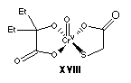 . The ligands bound to the chromium(V) center are the same for (XVI) and (XVIII), so it is not surprising that they result in the same signal in the EPR spectrum.
5.2. Summary
In the reactions discussed above, it is clear that in most cases, reduction of bis(2-ethyl-2-hydroxybutyrato)oxochromate(V) or bis(2-hydroxy-2-methylbutyrato)oxochromate(V) occurs before any appreciable ligand substitution on the chromium(V) center can take place. However, especially in non-aqueous solvents where reduction is retarded, reactions between the initial chromium(V) complex and dithiothreitol or thioglycolic acid resulted in ligand substitution, and the formation of mono- or bis-(thiol)chromium(V) complexes. Table 5-1 summarizes the EPR spectral data obtained in these studies.
Ghosh et al. (53) have studied the reduction of bis(2-ethyl-2-hydroxybutyrato)oxochromate(V) by the mercapto acids cysteine and thiolactic acid in acidic aqueous solvent. Based on kinetic data these authors have proposed a series of 1 e
- steps by which the starting chromium(V) complex is reduced:
Cr(V) + RSH
Æ Cr(IV) + RS• + H+ (k1) [5-2]
Cr(V) + RS•
Æ Cr(IV) + RS+ (k2) [5-3]
Cr(IV) + RSH
Æ Cr(III) + RS• + H+ (k3) [5-4]
Cr(IV) + RS•
Æ Cr(III) + RS+ (k4) [5-5]
and they have implicated a chromium(V)-thiolactate complex in the initial act of electron transfer (53). Initially,
[5-2] and [5-3] predominate, but as the level of chromium(IV) increases, the production of thiyl radical also increases; thiyl radical reacts preferentially with chromium(V) vs. chromium(IV) (k2 > k4), so autocatalysis is observed (53). The present study has shown that the reaction of bis(2-ethyl-2-hydroxybutyrato)oxochromate(V) with thioglycolic acid in non-aqueous solvent results in the formation of a chromium(V)-thioglycolate complex, and subsequent loss of the chromium(V) EPR signals. Reduction is retarded in non-aqueous solvent, so the chromium(V)-thioglycolate complex in this system is allowed to reach EPR-detectable levels. Also, the electronic spectra for the reaction with thioglycolic acid reveal the presence of chromium(III) species with a maximum at 444 nm at early times. Ghosh et al. (53) have attributed analogous absorbance maxima in their studies of reactions with thiolactic acid to a chromium(III) complex having a single S-bound ligand and five O-bound ligands. These data, as well as EPR evidence from the present study, are consistent with the formation of a chromium(V)(ehba)(thioglycolate) complex (XVIII) which is rapidly reduced to a chromium(III)(ehba)(thioglycolate) complex by the reaction steps outlined in equations [5-2] to [5-5].
Figure 5-1. EPR spectra of chromium(V) (A) and chromium(III) (B) complexes formed upon reaction of sodium bis(2-hydroxy-2-methylbutyrato)oxochromate(V) (50.0 mM chromium(V)) with
b-mercaptoethanol (50.0 mM; 1:1 thiol:Cr ratio). All reactions were performed at 25 °C in aqueous solution, initial pH 5.9. Reactions were initiated by addition of thiol, and aliquots (350 µL) of the reactions were taken at various times in quartz EPR tubes and quenched by immersion in liquid nitrogen. The initial chromium(V) EPR spectrum is not shown in panel (B) for clarity; no chromium(III) signals were observed in this region for stock chromium(V). EPR spectra were obtained as described in Figure 3-2.
placeholder for Figure 5-1
CrVBME290
Figure 5-2. EPR spectra of chromium(V) (A) and chromium(III) (B) complexes formed upon reaction of sodium bis(2-hydroxy-2-methylbutyrato)oxochromate(V) (5.0 mM chromium(V)) with
b-mercaptoethanol (5.0 mM; 1:1 thiol:Cr ratio) in 1:1 acetic acid:water solution and -15 °C. Reactions were initiated by addition of thiol, and aliquots (350 µL) of the reactions were taken at various times in quartz EPR tubes and quenched by immersion in liquid nitrogen. The initial chromium(V) EPR spectrum is not shown in panel (B) for clarity; no chromium(III) signals were observed in this region for stock chromium(V). EPR spectra were obtained as described in Figure 3-2.
placeholder for Figure 5-2
CrVBME295frozen
Figure 5-3. Solution EPR spectra of chromium(V) complexes formed upon reaction of sodium bis(2-hydroxy-2-methylbutyrato)oxochromate(V) (5.0 mM chromium(V)) with
b-mercaptoethanol (5.0 mM; 1:1 thiol:Cr ratio) in 1:1 acetic acid:water solution and -15 °C. Reactions were initiated by addition of thiol, and aliquots (50 µL) of the reactions were taken at various times in Pyrex capillary tubes and EPR spectra were obtained as described in Figure 3-7.
placeholder for Figure 5-3
CrVBME295RT
Figure 5-4. Solution EPR spectra of chromium(V) complexes formed upon reaction of sodium bis(2-hydroxy-2-methylbutyrato)oxochromate(V) (5.0 mM chromium(V)) with
b-mercaptoethanol (5.0 mM; 1:1 thiol:Cr ratio) in 1:1 acetic acid:water solution and -15 °C. Reactions were initiated by addition of thiol, and aliquots (50 µL) of the reactions were taken at various times in Pyrex capillary tubes and EPR spectra were obtained as described in Figure 3-7, with a modulation amplitude of 0.1 Gauss.
placeholder for Figure 5-4
CrVBME297aRT
Figure 5-5. Solution EPR spectra of chromium(V) complexes formed upon reaction of sodium bis(2-hydroxy-2-methylbutyrato)oxochromate(V) (5.0 mM chromium(V)) with
b-mercaptoethanol (5.0 mM; 1:1 thiol:Cr ratio) in 1:1 acetic acid:water solution with I = 1.5 M (maintained via KCl) and -15 °C. Reactions were initiated by addition of thiol, and aliquots (50 µL) of the reactions were taken at various times in Pyrex capillary tubes and EPR spectra were obtained as described in Figure 3-7, with a modulation amplitude of 0.1 Gauss.
placeholder for Figure 5-5
CrVBME297bRT
Figure 5-6. Solution EPR spectra of chromium(V) complexes formed upon reaction of sodium bis(2-hydroxy-2-methylbutyrato)oxochromate(V) (5.0 mM chromium(V)) with
b-mercaptoethanol (10.0 mM; 2:1 thiol:Cr ratio) in 1:1 acetic acid:water solution and -15 °C. Reactions were initiated by addition of thiol, and aliquots (50 µL) of the reactions were taken at various times in Pyrex capillary tubes and EPR spectra were obtained as described in Figure 3-7.
placeholder for Figure 5-6
CrVBME308RT
Figure 5-7. Solution EPR spectra of chromium(V) complexes formed upon reaction of sodium bis(2-hydroxy-2-methylbutyrato)oxochromate(V) (5.0 mM chromium(V)) with
b-mercaptoethanol (10.0 mM; 2:1 thiol:Cr ratio) in acetone at 25 °C. Reactions were initiated by addition of thiol, and aliquots (50 µL) of the reactions were taken at various times in Pyrex capillary tubes and EPR spectra were obtained as described in Figure 3-7. The inset to the figure shows the splitting of the 53Cr hyperfine satellite at high field; as the reaction proceeds this splitting becomes more prominent.
placeholder for Figure 5-7
CrVBME311
Figure 5-8. Solution EPR spectra of chromium(V) complexes formed upon reaction of sodium bis(2-hydroxy-2-methylbutyrato)oxochromate(V) (5.0 mM chromium(V)) with
b-mercaptoethanol (100.0 mM; 20:1 thiol:Cr ratio) in acetone at 25 °C. The insets to the figure show the splitting of the outer 53Cr satellites, and also a time-course for the g = 1.980 signal (values indicate mean + S.D. of two determinations). Reactions were initiated by addition of thiol, and aliquots (50 µL) of the reactions were taken at various times in Pyrex capillary tubes and EPR spectra were obtained as described in Figure 3-7.
placeholder for Figure 5-8
CrVBME312
Figure 5-9. Solution EPR spectra of chromium(V) complexes formed upon reaction of sodium bis(2-hydroxy-2-methylbutyrato)oxochromate(V) (5 mM chromium(V)) with
b-mercaptoethanol (500 mM; 100:1 thiol:Cr ratio) in acetone at 25 °C. (A) Main g = 1.980 signal and (B) blowup to show 53Cr hyperfine and splitting of 53Cr satellite at high field. Reactions were initiated by addition of thiol, and aliquots (50 µL) of the reactions were taken at various times in Pyrex capillary tubes and EPR spectra were obtained as described in Figure 3-7.
placeholder for Figure 5-9
CrVBME313
Figure 5-10. Solution EPR spectra of chromium(V) complexes formed upon reaction of potassium perchromate, K
3CrO8 (5.0 mM chromium(V)) with dithiothreitol (15.0 mM; 3:1 thiol:Cr ratio) in 0.2 N KOH (aqueous) at 25 °C. Reactions were initiated by addition of thiol, and aliquots (50 µL) of the reactions were taken at various times in Pyrex capillary tubes and EPR spectra were obtained as described in Figure 3-7.
placeholder for Figure 5-10
CrVDTT356
Figure 5-11. Solution EPR spectra of chromium(V) complexes formed upon reaction of sodium bis(2-ethyl-2-hydroxybutyrato)oxochromate(V) (5.0 mM) with dithiothreitol (10.0 mM; 2:1 thiol:Cr ratio) in 1:1 acetic acid:water solution at 25 °C. Reactions were initiated by addition of thiol, and aliquots (50 µL) of the reactions were taken at various times in Pyrex capillary tubes and EPR spectra were obtained as described in Figure 3-7.
placeholder for Figure 5-11
CrVdtt320
Figure 5-12. Solution EPR spectra of chromium(V) complexes formed upon reaction of sodium bis(2-hydroxy-2-methylbutyrato)oxochromate(V) (5.0 mM) with dithiothreitol (100.0 mM; 20:1 thiol:Cr ratio) in acetone at 25 °C. (A) Main g = 1.980 signal, and (B) blowup to show hyperfine and new g = 1.983 signal. Reactions were initiated by addition of thiol, and aliquots (50 µL) of the reactions were taken at various times in Pyrex capillary tubes and EPR spectra were obtained as described in Figure 3-7.
placeholder for Figure 5-12
CrVdtt316
Figure 5-13. (A) Solution EPR spectra of chromium(V) complexes formed upon reaction of sodium bis(2-ethyl-2-hydroxybutyrato)oxochromate(V) (5.0 mM) with dithiothreitol (100.0 mM; 20:1 thiol:Cr ratio) in acetonitrile at 25 °C. (B) Time-course of the main g = 1.980 signal. Values indicate mean + S.D. of two determinations. (C) Blowup of the spectra to show a
53Cr hyperfine structure. Reactions were initiated by addition of thiol, and aliquots (50 µL) of the reactions were taken at various times in Pyrex capillary tubes and EPR spectra were obtained as described in Figure 3-7.
placeholder for Figure 5-13
CrVdtt331
Figure 5-14. Solution EPR spectra of chromium(V) complexes formed upon reaction of sodium bis(2-ethyl-2-hydroxybutyrato)oxochromate(V) (5.0 mM) with dithiothreitol (25.0 mM; 5:1 thiol:Cr ratio) in dimethylformamide at 25 °C. (A) Main g = 1.980 signal, and (B) blowup to show hyperfine and new g = 1.983 signal. Reactions were initiated by addition of thiol, and aliquots (50 µL) of the reactions were taken at various times in Pyrex capillary tubes and EPR spectra were obtained as described in Figure 3-7.
placeholder for Figure 5-14
CrVdtt337
Figure 5-15. (A) Solution EPR spectra of chromium(V) complexes formed upon reaction of sodium bis(2-ethyl-2-hydroxybutyrato)oxochromate(V) (5.0 mM) with dithiothreitol (100.0 mM; 20:1 thiol:Cr ratio) in methanol at 25 °C. (B) Blowup to show hyperfine and new species' formation. Reactions were initiated by addition of thiol, and aliquots (50 µL) of the reactions were taken at various times in Pyrex capillary tubes and EPR spectra were obtained as described in Figure 3-7.
placeholder for Figure 5-15
CrVdtt333
Figure 5-16. (A) Solution EPR spectra of chromium(V) complexes formed upon reaction of sodium bis(2-ethyl-2-hydroxybutyrato)oxochromate(V) (5.0 mM) with dithiothreitol (25.0 mM; 5:1 thiol:Cr ratio) in methanol at 25 °C. (B) Blowup to show hyperfine and new species' formation. Reactions were initiated by addition of thiol, and aliquots (50 µL) of the reactions were taken at various times in Pyrex capillary tubes and EPR spectra were obtained as described in Figure 3-7.
placeholder for Figure 5-16
CrVdtt334
Figure 5-17. (A) Solution EPR spectra of chromium(V) complexes formed upon reaction of sodium bis(2-ethyl-2-hydroxybutyrato)oxochromate(V) (5.0 mM) with dithiothreitol (7.5 mM; 1.5:1 thiol:Cr ratio) in methanol at 25 °C. (B) Blowup to show hyperfine and new species' formation. Reactions were initiated by addition of thiol, and aliquots (50 µL) of the reactions were taken at various times in Pyrex capillary tubes and EPR spectra were obtained as described in Figure 3-7.
placeholder for Figure 5-17
CrVdtt335
Figure 5-18. Time course of the g = 1.980 EPR signal observed during the reaction of sodium bis(2-ethyl-2-hydroxybutyrato)oxochromate(V) (5.0 mM) with dithiothreitol (20, 5, or 1.5:1 thiol:Cr ratio) in methanol at 25 °C. Reactions were initiated by addition of thiol, and aliquots (50 µL) of the reactions were taken at various times in Pyrex capillary tubes and EPR spectra were obtained as described in Figure 3-7. Values represent mean + S.D. of two determinations. The inset to the figure displays the early time-points without error bars for clarity.
placeholder for Figure 5-18
CrVdtt333_5time
Figure 5-19. Comparison of the solution EPR spectra of sodium bis(2-ethyl-2-hydroxybutyrato)oxochromate(V) (5.0 mM) in various solvents (50% acetic acid, acetonitrile, N,N-dimethylformamide, or methanol) at 25 °C. Aliquots (50 µL) of the solutions were taken in Pyrex capillary tubes and EPR spectra were obtained as described in Figure 3-7.
placeholder for Figure 5-19
CrVrtcompare
Figure 5-20. Solution EPR spectra of chromium(V) complexes formed upon reaction of sodium dichromate (5.0 mM chromium(VI)) with dithiothreitol (25.0 mM; 5:1 thiol:Cr ratio) in methanol at 25 °C. Reactions were initiated by addition of thiol, and aliquots (50 µL) of the reactions were taken at various times in Pyrex capillary tubes and EPR spectra were obtained as described in Figure 3-7. Spectra shown are (A) 5.75 minutes and (B) 13.75 minutes after start of reaction. In (B) the number of scans averaged was higher resulting in an average signal between 8-15 minutes.
placeholder for Figure 5-20
CrVdtt336
Figure 5-21. Solution EPR spectra of chromium(V) complexes formed upon reaction of sodium bis(2-hydroxy-2-methylbutyrato)oxochromate(V) (5.0 mM) with glutathione (5.0 mM; 1:1 thiol:Cr ratio) in 1:1 acetic acid:water solution at 25 °C. Reactions were initiated by addition of thiol, and aliquots (50 µL) of the reactions were taken at various times in Pyrex capillary tubes and EPR spectra were obtained as described in Figure 3-7. (A) Full-sized spectra showing the main chromium(V) signal at g = 1.980, and (B) Blow-up to show hyperfine features.
placeholder for Figure 5-21. CrVgsh301
Figure 5-22. Solution EPR spectra of chromium(V) complexes formed upon reaction of sodium bis(2-hydroxy-2-methylbutyrato)oxochromate(V) (0.5 mM) with glutathione (5.0 mM; 10:1 thiol:Cr ratio) in 1:1 acetic acid:water solution at 25 °C. Reactions were initiated by addition of thiol, and aliquots (50 µL) of the reactions were taken at various times in Pyrex capillary tubes and EPR spectra were obtained as described in Figure 3-7. (A) Full-sized spectra showing the main chromium(V) signal at g = 1.980, and (B) Blow-up to show hyperfine features and new signal at g = 1.983.
placeholder for Figure 5-22. CrVgsh302
Figure 5-23. Solution EPR spectra of chromium(V) complexes formed upon reaction of sodium bis(2-hydroxy-2-methylbutyrato)oxochromate(V) (5.0 mM) with glutathione (15.0 mM; 3:1 thiol:Cr ratio) in 1:1 acetic acid:water solution at -15 °C. Reactions were initiated by addition of thiol, aliquots (50 µL) of the reactions were taken at various times in Pyrex capillary tubes and EPR spectra were obtained as described in Figure 3-7, with the temperature of the sample in the cavity held constant at -15 °C by means of a Bruker Variable Temperature apparatus. (A) Full-sized spectra showing the main chromium(V) signal at g = 1.980, and (B) Blow-up to show hyperfine features.
placeholder for Figure 5-23. CrVgsh309
Figure 5-24. Solution EPR spectra of chromium(V) complexes formed upon reaction of sodium bis(2-ethyl-2-hydroxybutyrato)oxochromate(V) (5.0 mM) with glutathione (100.0 mM; 20:1 thiol:Cr ratio) in acetone at 25 °C. Reactions were initiated by addition of thiol, and aliquots (50 µL) of the reactions were taken at various times in Pyrex capillary tubes and EPR spectra were obtained as described in Figure 3-7.
placeholder for Figure 5-24. CrVgsh323
Figure 5-25. Solution EPR spectra of chromium(V) complexes formed upon reaction of sodium bis(2-ethyl-2-hydroxybutyrato)oxochromate(V) (5.0 mM) with glutathione (100.0 mM; 20:1 thiol:Cr ratio) in N,N-dimethylformamide at 25 °C. Reactions were initiated by addition of thiol, and aliquots (50 µL) of the reactions were taken at various times in Pyrex capillary tubes and EPR spectra were obtained as described in Figure 3-7. (A) Full-sized spectra showing the main chromium(V) signal at g = 1.980, and (B) Blow-up to show hyperfine features.
placeholder for Figure 5-25. CrVgsh338
Figure 5-26. Solution EPR spectra of chromium(V) complexes formed upon reaction of sodium bis(2-ethyl-2-hydroxybutyrato)oxochromate(V) (5.0 mM) with oxidized glutathione (50.0 mM; 10:1 disulfide:Cr ratio) in aqueous solution at 25 °C (initial pH = 3.8). Reactions were initiated by addition of disulfide, and aliquots (50 µL) of the reactions were taken at various times in Pyrex capillary tubes and EPR spectra were obtained as described in Figure 3-7. (A) Full-sized spectra showing the main chromium(V) signal at g = 1.980, and (B) Blow-up to show hyperfine features.
placeholder for Figure 5-26.
CrVgssg293
Figure 5-27. Frozen solution (77K) EPR spectra of chromium(V) complexes formed upon reaction of sodium bis(2-ethyl-2-hydroxybutyrato)oxochromate(V) (5.0 mM) with oxidized glutathione (50.0 mM; 10:1 disulfide:Cr ratio) in aqueous solution at 25 °C (initial pH = 3.8). Reactions were initiated by addition of disulfide, and aliquots (350 µL) of the reactions were taken at various times in quartz EPR tubes and quenched by immersion in liquid nitrogen. EPR spectra were obtained as described in Figure 3-2. (A) Full-sized spectra showing the main chromium(V) signal at g = 1.980; Blow-up to show g = 1.980 (C) and g = 5 (B) features.
placeholder for Figure 5-27.
CrVgssg293B
Figure 5-28. Solution EPR spectra of chromium(V) complexes formed upon reaction of sodium bis(2-hydroxy-2-methylbutyrato)oxochromate(V) (5.0 mM) with glycine (5.0, 50.0, or 500.0 mM; 1:1,10, or 100 Cr:glycine ratio) in 1:1 acetic acid:water solution at 25 °C. Reactions were initiated by addition of glycine, and aliquots (50 µL) of the reactions were taken at various times in Pyrex capillary tubes and EPR spectra were obtained as described in Figure 3-7.
placeholder for Figure 5-28.
CrVgly298
Figure 5-29. Solution EPR spectra of chromium(V) complexes formed upon reaction of sodium bis(2-hydroxy-2-methylbutyrato)oxochromate(V) (4.0 mM) with oxalic acid (200.0 mM; 1:50 Cr:oxalic acid ratio) in 1:1 acetic acid:water solution at 25 °C. Reactions were initiated by addition of oxalic acid, and aliquots (50 µL) of the reactions were taken at 1.3 minutes in Pyrex capillary tubes and EPR spectra were obtained as described in Figure 3-7.
placeholder for Figure 5-29.
CrVox303
Figure 5-30. Solution EPR spectra of chromium(V) complexes formed upon reaction of sodium bis(2-ethyl-2-hydroxybutyrato)oxochromate(V) (4.0 mM) with oxalic acid (200.0 mM; 1:50 Cr:oxalic acid ratio) in 1:1 acetic acid:water solution at 25 °C. Reactions were initiated by addition of oxalic acid, and aliquots (50 µL) of the reactions were taken at various times in Pyrex capillary tubes and EPR spectra were obtained as described in Figure 3-7.
placeholder for Figure 5-30.
CrVox318
Figure 5-31. Solution EPR spectra of chromium(V) complexes formed upon reaction of sodium bis(2-hydroxy-2-methylbutyrato)oxochromate(V) (4.0 mM) with oxalic acid (200.0 mM; 1:50 Cr:oxalic acid ratio) in acetone at 25 °C. Reactions were initiated by addition of oxalic acid, and aliquots (50 µL) of the reactions were taken at various times in Pyrex capillary tubes and EPR spectra were obtained as described in Figure 3-7.
placeholder for Figure 5-31.
CrVox315
Figure 5-32. Solution EPR spectra of chromium(V) complexes formed upon reaction of sodium bis(2-ethyl-2-hydroxybutyrato)oxochromate(V) (4.0 mM) with oxalic acid (200.0 mM; 1:50 Cr:oxalic acid ratio) in acetone at 25 °C. Reactions were initiated by addition of oxalic acid, and aliquots (50 µL) of the reactions were taken at various times in Pyrex capillary tubes and EPR spectra were obtained as described in Figure 3-7.
placeholder for Figure 5-32.
CrVox319
Figure 5-33. Solution EPR spectra of chromium(V) complexes formed upon reaction of sodium bis(2-ethyl-2-hydroxybutyrato)oxochromate(V) (5.0 mM) with oxalic acid (1.0 M; 1:200 Cr:oxalic acid ratio) in acetone at 25 °C. Reactions were initiated by addition of oxalic acid, and aliquots (50 µL) of the reactions were taken at various times in Pyrex capillary tubes and EPR spectra were obtained as described in Figure 3-7.
placeholder for Figure 5-33.
CrVox322
Figure 5-34. Solution EPR spectra of chromium(V) complexes formed upon reaction of sodium bis(2-ethyl-2-hydroxybutyrato)oxochromate(V) (5.0 mM) with ascorbic acid (2.5 mM; 1:0.5 Cr:ascorbic acid ratio) in 1:1 acetic acid:water solution at 25 °C. Reactions were initiated by addition of ascorbic acid, and aliquots (50 µL) of the reactions were taken at various times in Pyrex capillary tubes and EPR spectra were obtained as described in Figure 3-7.
placeholder for Figure 5-34.
crvasc328a
Figure 5-35. Solution EPR spectra of chromium(V) complexes formed upon reaction of sodium bis(2-ethyl-2-hydroxybutyrato)oxochromate(V) (5.0 mM) with ascorbic acid (100.0 mM; 1:20 Cr:ascorbic acid ratio) in acetone at 25 °C. Reactions were initiated by addition of ascorbic acid, and aliquots (50 µL) of the reactions were taken at various times in Pyrex capillary tubes and EPR spectra were obtained as described in Figure 3-7.
placeholder for Figure 5-35.
crvasc327a
Figure 5-36. Solution EPR spectra of chromium(V) complexes formed upon reaction of sodium bis(2-ethyl-2-hydroxybutyrato)oxochromate(V) (5.0 mM) with thioglycolic acid (2.5 mM; 1:0.5 Cr:thioglycolic acid ratio) in 1:1 acetic acid:water solution at 25 °C. Reactions were initiated by addition of thiol, and aliquots (50 µL) of the reactions were taken at various times in Pyrex capillary tubes and EPR spectra were obtained as described in Figure 3-7.
placeholder for Figure 5-36.
crvtga329a
Figure 5-37. Solution EPR spectra of chromium(V) complexes formed upon reaction of sodium bis(2-ethyl-2-hydroxybutyrato)oxochromate(V) (5.0 mM) with thioglycolic acid (5.0 mM; 1:1 Cr:thioglycolic acid ratio) in 1:1 acetic acid:water solution at 25 °C. Reactions were initiated by addition of thiol, and aliquots (50 µL) of the reactions were taken at various times in Pyrex capillary tubes and EPR spectra were obtained as described in Figure 3-7.
placeholder for Figure 5-37.
crvtga329b
Figure 5-38. Solution EPR spectra of chromium(V) complexes formed upon reaction of sodium bis(2-ethyl-2-hydroxybutyrato)oxochromate(V) (5.0 mM) with thioglycolic acid (100.0 mM; 1:20 Cr:thioglycolic acid ratio) in acetone at 25 °C. Reactions were initiated by addition of thiol, and aliquots (50 µL) of the reactions were taken at various times in Pyrex capillary tubes and EPR spectra were obtained as described in Figure 3-7.
placeholder for Figure 5-38.
crvtga326a
Figure 5-39. Solution EPR spectra of chromium(V) complexes formed upon reaction of sodium bis(2-ethyl-2-hydroxybutyrato)oxochromate(V) (5.0 mM) with thioglycolic acid (100.0 mM; 1:20 Cr:thioglycolic acid ratio) in acetonitrile at 25 °C. Reactions were initiated by addition of thiol, and aliquots (50 µL) of the reactions were taken at various times in Pyrex capillary tubes and EPR spectra were obtained as described in Figure 3-7.
placeholder for Figure 5-39.
crvtga330a
Figure 5-40. Solution EPR spectra of chromium(V) complexes formed upon reaction of sodium bis(2-ethyl-2-hydroxybutyrato)oxochromate(V) (5.0 mM) with thioglycolic acid (100.0 mM; 1:20 Cr:thioglycolic acid ratio) in N,N-dimethylformamide at 25 °C. Reactions were initiated by addition of thiol, and aliquots (50 µL) of the reactions were taken at various times in Pyrex capillary tubes and EPR spectra were obtained as described in Figure 3-7.
placeholder for Figure 5-40.
crvtga339a
Figure 5-41. Solution EPR spectra of chromium(V) complexes formed upon reaction of sodium bis(2-ethyl-2-hydroxybutyrato)oxochromate(V) (5.0 mM) with thioglycolic acid (100.0 mM; 1:20 Cr:thioglycolic acid ratio) in methanol at 25 °C. Reactions were initiated by addition of thiol, and aliquots (50 µL) of the reactions were taken at various times in Pyrex capillary tubes and EPR spectra were obtained as described in Figure 3-7.
placeholder for Figure 5-41.
crvtga332a
Figure 5-42. Electronic spectra of the reaction of sodium bis(2-ethyl-2-hydroxybutyrato)oxochromate(V) (5.0 mM) with thioglycolic acid (100.0 mM; 1:20 Cr:thioglycolic acid ratio) in methanol at 25 °C. Reactions were initiated by addition of thiol, and spectra were obtained using a Perkin-Elmer Lambda-9 dual beam spectrophotometer using 1 cm quartz cells. The scan rate was 2.5 minutes per spectrum and a slit width of 2 nm was employed.
placeholder for Figure 5-42.
crvtga346
Table 5-1. EPR spectral parameters for the chromium(V) signals observed upon reaction of chromium(V) complexes with reducing agents in various solvents.
table 5-1 holder, page 2
table 5-1 holder, page 3