 .
.6. Summary of Results and Implications for Chromium(VI) Carcinogenesis
The overall goal of this research has been to illuminate the possible fate of chromium(VI) and chromium(V) in biological systems, by studying in vitro reactions of chromium(VI) and chromium(V) with low molecular weight biological and model reductants, as a means of more fully understanding the processes involved in the genotoxicity of chromium. Toward this end, the main focus of this research has been the study of chromium(V) complexes and other reactive intermediates formed during reactions of chromium(VI) and chromium(V) with thiols and other reducing agents such as ascorbate.
It is probable that chromium(VI) must be "activated" as the first step in its carcinogenic process, since in vitro studies have shown that chromium(VI) does not interact with DNA in the absence of a reducing system (13). Reactive intermediates formed upon reduction of chromium(VI) in vivo would likely consist of chromium(V), chromium(IV), and radical species such as hydroxyl and thiyl radicals (19). These reactive intermediates would then attack DNA (75) causing DNA damage, and ultimately, tumors. A number of in vitro studies have shown that chromium(V) is formed when chromium(VI) is reacted with cellular constituents such as hydrogen peroxide (22), ascorbic acid (20), and glutathione (70, 76, 77). Furthermore, recent studies have shown that chromium(V) is formed when cultured Chinese hamster V-79 cells are exposed to chromium(VI) (78). Sugiyama et al. (78-80) have reported a differential effect on chromium(VI)-induced DNA damage when Chinese hamster V-79 cells are pre-incubated with either vitamin E or vitamin B
2. The authors have shown that pre-treatment with vitamin B2 resulted in an increase in the levels of chromium(V) observed, as well as in increase in the formation of DNA strand breaks (79, 80). However, pre-treatment with vitamin E resulted in a decrease in the levels of chromium(V) and a decrease in the formation of DNA strand breaks (78, 80). It is suggested that the differential effects of vitamins B2 and E on the formation of chromium(VI)-induced DNA strand-breaks may be due to modification of chromium(V) formation in cells (80). Furthermore, Sugiyama et al. (81) have reported that when Chinese hamster V-79 cells are pre-incubated with ascorbic acid the number of DNA-protein cross-links produced by chromium(VI) are increased, while the levels of chromium(VI)-induced alkali-labile sites decreased. Pre-treatment with ascorbate also reduced the levels of chromium(V) observed in this system (81). Aiyar et al. (82) have shown that two chromium(V) complexes, glutathione thiyl radical, and chromium-DNA adducts are formed by the reaction of chromium(VI) with glutathione, but little DNA strand breaks or 8-hydroxydeoxyguanosine adducts were formed. Reactions between chromium(VI) and hydrogen peroxide, on the other hand, led to DNA strand breaks and 8-hydroxydeoxyguanosine adducts in the absence of detectable chromium(V) or chromium-DNA adducts (82). Borges and Wetterhahn (14) have shown that in vitro reduction of chromium(VI) by glutathione or cysteine leads to glutathione-chromium-DNA and (cysteine)2-4-chromium-DNA binding (14). A recent study by Kortenkamp et al. (83) has shown that significant chromium-DNA binding occurs when isolated cell nuclei are incubated with chromium(VI) in the presence of glutathione, but very limited binding occurs in the absence of glutathione; this indicates that chromium(VI) must be reduced (presumably to chromium(V)) in order to bind to cell nuclei. Farrell et al. have shown (84) that DNA strand-cleavage occurs when plasmid DNA is incubated with the chromium(V) compound [CrV(ehba)2O]-. These authors have also shown that [CrV(ehba)2O]-, [CrV(hmba)2O]-, and Cr(VI) (as dichromate) have equivalent mutagenic activity, based on the number of revertants of a histidine auxotroph of Salmonella typhimurium that is sensitive to chromium(VI) mutagenesis (84). An in vivo study in our laboratory has shown that chromium(V) is formed in red blood cells when chick embryos are exposed to chromium(VI) (85). This is further evidence implicating chromium(V) as the "ultimate" carcinogen in vivo. The present studies have expanded previous studies and have focused on such chromium(V) and radical species, in an attempt to further our understanding of the mechanism by which chromium(VI) compounds can lead to tumors.Reduction of chromium(VI) by any of the thiols
b-mercaptoethanol, dithiothreitol, glutathione, or cysteine leads to the formation of multiple chromium(V) species, as well as thiyl or hydroxyl radicals. The level of chromium(V) formed in reactions with these thiols is related to the level of chromium-DNA binding observed in reactions performed in the presence of DNA (63). Reactions involving b-mercaptoethanol and dithiothreitol produced large amounts of chromium(V) and a high level of chromium-DNA binding, while cysteine and glutathione effected very low levels of chromium(V) and low levels of chromium-DNA binding. The highest level of chromium(III) formation was observed in reactions involving cysteine. However, cysteine effected the lowest chromium-DNA binding; the converse was true for b-mercaptoethanol. Thus, the levels of chromium bound to DNA in the presence of these thiols does not correlate with production of EPR-detectable chromium(III). These data are particularly intriguing, and provide convincing evidence that chromium(V) species serve as the "reactive" form of chromium that attacks DNA and leads to DNA damage. Studies done in our laboratory have shown that chromium(V) is formed in vivo (85), and thus it is proposed that chromium(V) is the ultimate reactive, DNA-damaging agent in vivo. Also observed in the present studies was the formation of significant amounts of thiyl radical, especially in reactions involving b-mercaptoethanol. Levels of chromium(V) formed in the presence of agents capable of trapping these thiyl radicals (such as DNA) were dramatically increased. Since thiyl radical is observed in reactions involving most of the thiols studied, and DNA has been shown to effectively "trap" such radicals, thiyl radical may also play a role in inducing DNA damage. Reduction of chromium(VI) by cysteine or cysteamine produced low levels of hydroxyl radical, which can also cause DNA damage. Kortenkamp et al. (86) have reported that reactions of chromium(VI) and glutathione performed in the presence of supercoiled circular bacteriophage PM2 DNA resulted in DNA strand breaks, in contrast to the results reported by Aiyar et al. (82) who have shown that very little strand breaks occur upon incubation of DNA with chromium(VI) and glutathione. However, Kortenkamp et al. (86) did not treat their buffer solutions to remove trace levels of iron, which is capable of generating hydroxyl radicals that can cause DNA strand breakage. In a subsequent study, Kortenkamp et al. (68) reported that addition of iron to their system did result in increasing levels of DNA strand breaks. They also report that the levels of DNA strand breaks were dependent upon both the ratio of chromium(VI):glutathione and the absolute concentration of the reactants (68). Thus, the data reported by Kortenkamp et al. (68) suggests that at the levels of chromium(VI) used by Aiyar et al. (82), any hydroxyl radical that is formed would be scavenged by the high levels of glutathione (27 mM), thus preventing the formation of DNA strand breaks. Jones et al. (87) have shown that hydroxyl radicals (•OH) can be formed by a chromium(V) intermediate isolated from the reaction of chromium(VI) with glutathione, and have proposed that the generation of hydroxyl radical involves the glutathione thiyl radical (RS•) [6-1] to [6-3]:RS• + RSH + O
2 Æ RSSR + O2•- + H+ [6-1]O
2•- + O2•- + 2H+ Æ H2O2 + O2 [6-2]M
(n-1)+ + H2O2 Æ Mn+ + •OH + OH- [6-3]In this mechanism, thiyl radical (RS•) reacts with excess glutathione (RSH) to form superoxide (O
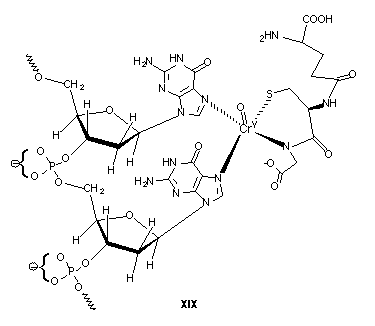
2•-), which in turn can generate hydrogen peroxide (H2O2). Hydroxyl radicals (•OH) can then be formed via Fenton chemistry where M can be iron, chromium, or another redox active metal.The present studies have also allowed the structures of the chromium(V) complexes formed during the reaction of chromium(VI) with thiols to be postulated, and all involve an axial geometry about the chromium(V) center. The observed g-values of these chromium(V) complexes have given information about the ligands bound to the chromium(V) center. In the in vivo study by Liebross and Wetterhahn (85) the g-values of the observed chromium(V) complexes (g = 1.981, 1.992) compare favorably to the g-values of chromium(V) complexes observed for reactions of chromium(VI) with glutathione (g = 1.987, 1.998) discussed in chapters 3 and 4. The g = 1.987, 1.998 features have been assigned in this study to mono(glutathione)chromium(V) (V) and bis(glutathione)chromium(V) (VI) complexes, respectively. It is thus probable that the chromium(V) signals observed in the in vivo studies are due to glutathione-chromium(V) complexes (85). Since the formation and decay of the chromium(V) species observed in vivo was relatively rapid (after a short lag-time necessary for uptake of chromium(VI)), and only a small amount of g = 1.992 signal was observed, it is postulated that the mono(glutathione)chromium(V) complex (V) is formed upon exposure to chromium(VI). The present studies have shown that chromium(V) levels are related to levels of chromium-DNA binding, so it is possible that in the in vivo system of Liebross and Wetterhahn (85) a mono-(glutathione)chromium(V) complex (V) is the main reactive chromium(VI) metabolite, and this complex (V) reacts with DNA, forming a (glutathione)Cr
V(DNA) complex, (XIX):.
Complex (XIX) involves coordination of the mono(glutathione)chromium(V) complex (V) to the N7 positions of adjacent guanine residues on DNA. Borges and Wetterhahn (14) have shown that thiol-chromium-DNA adducts formed upon incubation of chromium(VI) and glutathione with DNA form preferentially at guanine bases. Many transition metal complexes can coordinate with nitrogen lone pair electrons on DNA bases, forming covalent bonds (88, 89). The platinum anticancer drug, cis-diamminedichloroplatinum(II), forms an intrastrand cross-link through the N7 atoms of adjacent guanines (90). Thus, it is likely that any chromium-DNA complexes formed would involve ligation at the N7 positions of adjacent guanines. The g = 1.992 signal observed by Liebross and Wetterhahn (85) is consistent with increasing covalency when going from S, N, O, O ligation in (V) (g = 1.987) to S, N, N, N ligation in (XIX). Farrell et al. (84) have shown that the presence of excess 2-ethyl-2-hydroxybutanoato ligand diminishes the DNA cleavage reaction of [Cr
V(ehba)2O]-. The authors have postulated that a chromium(V)-DNA complex would form prior to DNA strand cleavage, thus the presence of excess 2-ethyl-2-hydroxybutanoato ligand must favor [CrV(ehba)2O]-. Loss of ligand would be required prior to DNA binding and thus the chromium(V) complex cannot bind to DNA and cause strand breaks in the presence of excess ligand (84). This is further evidence that chromium(V) is the main reactive species involved in DNA damage.Most of the thiols used in the present study have been shown to produce chromium(V) and thiyl radicals upon incubation with chromium(VI), and in most cases the formation of mono-(thiol)chromium(V) complexes and subsequent formation of bis-(thiol)chromium(V) complexes is consistent with the EPR data observed. It is clear from our work and the work in other laboratories that there are numerous parallel routes of chromium(VI) metabolism leading to DNA damage. These pathways can be divided into two main categories: (A) Formation of chromium(V) species leading to chromium-DNA binding and (B) DNA-radical adducts arising from reactive radical species formed during the metabolism of chromium(VI) and chromium(V). The binding of chromium to DNA (14, 63, 82) would most likely lead to ternary thiol-chromium-DNA complexes such as (XIX) (and ultimately analogous ternary chromium(III) complexes). Alternatively, the binding of chromium to DNA may take place at the phosphate backbone, leading to oxidative cleavage (84, 86). It is possible that both types of DNA-binding occur, however the chromium(V) species observed by Liebross and Wetterhahn (85) is consistent with the complex (XIX) and the results of the present study. Hamilton and Wetterhahn (8) have shown that treatment of 14-day chick embryos with chromium(VI) lead to the formation of significant DNA strand breaks in red blood cells 4 hours after treatment, and very low levels of DNA cross-links. However, the chromium(VI) dose used by Hamilton and Wetterhahn (8) was much lower (0.1 mmol/kg body weight sodium dichromate) than that used by Liebross and Wetterhahn (0.6 mmol/kg sodium dichromate) (85). At lower chromium(VI) doses DNA strand breaks but no chromium(V) is observed in the system of Liebross and Wetterhahn (91). Thus, the chromium(VI) dosage affects the overall metabolism of chromate in this system. The chromium(V) EPR signal observed in chick embryo red blood cells was very short-lived, indicating that the chromium(V) species is reactive. If complex (XIX) is formed, it is possible that upon further reduction to chromium(IV) or chromium(III) thiyl radical is generated. Any thiyl radical thus generated in close proximity to DNA could subsequently attack DNA leading to the observed strand breaks. The formation of DNA-radical adducts can occur via hydroxyl-radical or thiyl radical attack of DNA; both radicals can be formed during the metabolism of chromium(VI) and chromium(V), as shown by the present study and previous work (79, 82, 92, 93)
As in vivo studies of the metabolism of chromium(VI) in more systems are performed, it is expected that chromium(V) species will be observed, and the present studies will allow the structures of such species to be postulated. This information is crucial for understanding the mechanisms by which chromium(VI) compounds lead to cancer.