Structure of CuB in the Binuclear Heme-Copper Center of the Cytochrome aa3-Type Quinol Oxidase from Bacillus subtilis : An ENDOR and EXAFS Study
Yang C. Fann ¤; Ishak Ahmed **; Ninian J. Blackburn #; John S. Boswell #; Marina L. Verkhovskaya **; Brian M. Hoffman ¤; and Mårten Wikström * *** Address correspondence to this author at Department of Medical Chemistry,P.O. Box 8, 00014 University of Helsinki, Helsinki, Finland.
# Department of Chemistry, Biochemistry and Molecular Biology,Oregon Graduate Institute of Science and Technology, Portland, Oregon 97291
¤ Department of Chemistry, Northwestern University, Evanston, Illinois60208-3113
** Helsinki Bioenergetics Group, Institute of Biomedical Sciences,Department of Medical Chemistry, 00014 University of Helsinki, Helsinki,Finland
Received April 25, 1995; Revised Manuscript Received June 14, 1995
This work was supported by grants from the Sigrid Jusèlius Foundation,the Academy of Finland (M.R.C.), the University of Helsinki (to M.W.), and theNational Institutes of Health (HL 13531 to B.M.H. and NS 27583 to N.J.B.).
ABSTRACT:
We have studied the structure of the CuB site in the binuclear heme-copper center of the fully oxidized form of the quinol-oxidizing cytochrome aa3-600 from Bacillus subtilis by EXAFS and ENDOR spectroscopy. This enzyme is member of the large superfamily of heme-copper respiratory oxidases,which catalyze the reduction of dioxygen to water and link it to translocation of protons across the bacterial or mitochondrial membrane. The EXAFS of the CuB site strongly suggests tetragonal coordination by two or three histidines with one or two O/N donor ligands. There are some indications that a Cl - ion might fractionally occupy substitution-labile sites, although the majority of enzyme molecules did not contain any heavy (second row) scatterers, indicative of a Cl- (or S) bridge between the heme iron and CuB [cf. Powers, L., et al. (1994)Biochim. Biophys. Acta 1183, 504-512]. Proton ENDOR spectroscopy of the CuB site in 1H2O and 2H2O media showed evidence of an oxygenous copper ligand with an exchangeable proton. 14N ENDOR revealed three inequivalent nitrogenous ligands with hyperfine coupling constants consistent with histidines. Together, these results strongly suggest that the fully oxidized enzyme has alow-symmetry, tetragonal CuB site with three histidine nitrogens and one oxygen as ligands, the latter with an exchangeable proton(s). The identity and assignment of these ligands are discussed.
The respiratory heme-copper oxidases constitute a superfamily of proton-translocating terminal oxidases in both prokaryotic organisms and the mitochondria of eukaryotic cells (see Saraste, 1990; Hosler et al., 1993;Calhoun et al., 1994). The most widely studied enzyme is the cytochrome c oxidase of mammalian mitochondria, but its 13-subunit composition and nonamenability to mutagenesis have hampered progress in its structural characterization. On the other hand, a number of bacterial heme-copper oxidases contain only three subunits, which are homologous to the mtDNA-encoded large subunits in the mitochondrial enzyme and exhibit catalytic characteristics similar to the latter.
The cytochrome c oxidases contain three redox centers, CuA, 1 heme Fea, and the binuclear, dioxygen-binding heme Fea3-CuB center, and utilizecytochrome c as the electron donor. A second major branch of this enzyme familyuses ubiquinol or menaquinol as the reducing substrate, and in these cases theCuA center is absent. Such enzymes are the cytochrome bo3 of Escherichia coli(Puustinen et al., 1991, 1992; Minghetti et al., 1992; Hosler et al., 1993;Calhoun et al., 1994) and cytochrome aa3-600 of Bacillus subtilis (Lauraeus etal., 1991, 1992; Santana et al., 1992), which is the subject of the presentstudy. Many bacteria can be induced to produce a certain terminal oxidase whengrown under specified conditions. Thus, B. subtilis can be induced to produce acytochrome caa3-type cytochrome c oxidase in addition to cytochrome aa3-600,when grown aerobically on a nonfermentable carbon source, whereas cytochromeaa3-600, apparently the constitutive respiratory oxidase, is virtually the onlyone expressed during logarithmic growth on a fermentable substrate (Lauraeus etal., 1991).
There is remarkable sequence homology between subunits I of all members of thesuperfamily, and it is established that the binuclear heme Fea3-CuB dioxygenreduction site resides within this subunit, together with the low-spin hemeFea, while the CuA center is located in subunit II. Spectroscopic andsite-directed mutagenesis, as well as three-dimensional modeling, has led to aworking model (Hosler et al., 1993) for the basic structure of the Fea3-CuBcenter and its connectivity to Fea. Figure 1 shows one version of thismodel in which the distance between the heme edges is about 11 Å toaccount for the measured rate of heme-heme electron transfer (Verkhovsky etal., 1992).
According to this model, the metal centers of subunit I are coordinated byfully conserved histidine residues: Fea is coordinated to His102 fromtransmembrane helix II and to His421 on one side of helix X. His419 on theopposite side of helix X is the proximal ligand of Fea3, the distal side facingthe CuB site. CuB is coordinated to His333 and His334 of the putativetransmembrane helix VII, and possibly to His284 in helix VI as well. Recentmutagenesis studies have also suggested that Tyr288 (in helix VI) might be aligand to CuB in certain states of the enzyme, since its mutation to Phedestroys the CO-binding capacity at the reduced CuB site (Thomas et al., 1994).[Ref. 2]
The binuclear Fea3-CuB center is the site of dioxygen chemistry in all of theseenzymes. Moreover, it seems clear that this site is also intimately involved inthe proton-translocating function (Wikström, 1989), and several differentmodels of proton-translocating mechanisms have been proposed, all of which havethe Fea3-CuB center structure in common (Chance & Powers, 1985; Mitchell,1987, 1988; Wikström, 1988; Rousseau et al., 1993; Woodruff, 1993; Babcock& Wikström, 1992; Wikström et al., 1994; Morgan et al., 1994;Rich, 1995). From this it is clear that the structural information of thebinuclear site is of great importance in order to gain an understanding of theproton translocation mechanism, as well as of the catalytic mechanism ofdioxygen reduction.
The two metals of the binuclear Fea3-CuB center are within 5 Å from oneanother in the oxidized enzyme and form a normally EPR-undetectablespin-coupled pair (van Gelder & Beinert, 1969). The coupling is thought tobe mediated by a bridging ligand, the nature of which is still under debate.EPR signals are observed from the binuclear center when either of the twometals is reduced or when the magnetic coupling has been broken by other means.In redox equilibrium titrations, CuB is always reduced before Fea3, yieldinghigh-spin ferric signals from the latter, with CuB remaining EPR-invisible (seeWikström et al., 1981). However, intermediate states have been trappedduring turnover of the enzyme that show rhombic EPR signals from cupric CuB(Reinhammar et al., 1980; Karlsson et al., 1981). An ENDOR study of such atrapped state suggested that CuB may have three nitrogenous (histidine) ligandsand a fourth unknown ligand (Cline et al., 1983). The latter may be theputative bridging ligand in the oxidized state of the center or another(solvent) molecule replacing it. EPR studies have also demonstrated that awater or hydroxide molecule derived from O2 is coordinated to CuB in a trappedintermediate state of the catalytic cycle (Hansson et al., 1982).
Most EXAFS studies on the oxidized binuclear site reported to date have beendone on the mitochondrial enzyme and have suggested two or three (Powers etal., 1981; Li et al., 1987) nitrogenous ligands of CuB, plus one S/Cl ligandpresumed to bridge CuB with Fea3. A preliminary Fe EXAFS report by Scott et al.(1988) indicated that the putative bridging ligand was lost in chloride-freeconditions, but to our knowledge there has been no followup on this issue.Earlier EXAFS work was hampered by the fact that the mitochondrial cytochrome coxidase also contains a CuA site, which, by itself, is now known to contain twocopper ions (Lappalainen et al., 1993). For this reason, EXAFS studies of thestructurally homologous quinol oxidases appear more promising, since theseenzymes lack the CuA center and its ligands altogether. Recently, Powers et al.(1994) studied the quinol-oxidizing cytochrome aa3-600 from B. subtilis andreported EXAFS data very similar to those reported previously with themitochondrial enzyme. A heavy S/Cl scatterer was observed bridging the metals,the identity of which was considered to be chloride since no conservedcysteines or methionines could conceivably provide a sulfur and since noacid-labile sulfur was detected.
Here, we have continued the study of the structure of the CuB site of the B.subtilis cytochrome aa3-600 by both EXAFS and ENDOR techniques, with the aim ofresolving the long-standing problem of the identity of the bridging ligand andthe putative role of Cl - as such a ligand, as well as exploring thecoordination sphere of CuB more generally.
MATERIALS AND METHODS
Enzyme Samples and Chloride Determination.Cytochrome aa3-600 from B. subtiliswas isolated and purified as described previously (Lauraeus et al., 1991). Theenzyme concentration was determined from the dithionite-reduced minusair-oxidized optical difference spectrum at the wavelength couple 600-626 nm,using a millimolar absorptivity of 26.4 cm - 1 for the two-heme enzyme unit(Lauraeus et al., 1992).
Samples for ENDOR spectroscopy were prepared as follows: a 0.1 mM suspensionof cytochrome aa3-600 (in 50 mM Tris, 150 mM NaCl, and 0.5% Triton X-100, pH 8)was diluted 4 times in buffer containing 100 mM glycine and 150 mM NaCl (pH8.8). This was then concentrated to the original volume using AmiconCentricon-100 microconcentrators. This process of dilution and subsequentconcentration was repeated three times to ensure that the final sample was atpH 8.8. For samples in heavy water, the enzyme dilution was done in the sameglycine buffer as before, except that the buffer was made up in 2H2O at pD 8.8(pH-meter reading 8.4), and concentrated as before. The procedure was repeatedthree times to ensure that the 2H2O content exceeded 90%.
Samples for EXAFS spectroscopy were prepared as follows: 2% (w/v) potassiumcholate was added to about 35 mM enzyme in 50 mM Tris, 150 mM NaCl, and 0.3%Triton X-100 (pH 8.0). This suspension was 50% saturated with ammonium sulfateby adding saturated, neutralized ammonium sulfate solution to precipitate theenzyme. The precipitate was dissolved in buffer containing 50 mM Tris, 150 mMNaCl, 0.1% Triton X-100, and 10% glycerol (pH 8.0; the concentration ofcytochrome aa3-600 was about 0.35 mM in this stock solution).
To prepare the high-pH sample (pH 8.8), about 0.15 mL of stock enzyme solutionwas diluted 4 times in buffer containing 100 mM glycine, 150 mM NaCl, and 20%(v/v) glycerol (pH 8.8). This was then concentrated as before, and the processof dilution and concentration was repeated three times to ensure that the pHwas 8.8 in the final preparation.
The chloride-free samples were prepared as follows: 0.2 mM stock enzymesolution was dialyzed overnight against 0.5 L of Cl-free buffer (50 mM Tris, 75mM Na2SO4, 0.1% Triton X-100, and 20% (v/v) glycerol, pH 8.0). The externalbuffer was changed and dialysis continued for 5 h against 0.5 L of new buffer.The volume of the sample increased upon dialysis and was concentrated as beforeto a final concentration between 0.2 and 0.3 mM. The concentration of Cl - inthe enzyme suspension was determined using a Spectroquant kit (Merck Cl -14755) and was found to be <5 mM. This Cl - assay is based on the reactionof chloride with mercuric thiocyanate in the presence of ferric ions to formmercuric chloride, chloromercurate(II) anion, and orange-red ferricthiocyanate. Since salts and sugars influence the color, a calibration curvehad to be made with fresh dialysis medium.
In an attempt to determine Cl - that might be tightly bound to the enzyme, thedialysis bag was boiled in alkaline conditions for 10 min after completion ofdialysis, the suspension was acidified, and the protein was sedimented andfiltered on a Centricon column. Then Cl - was determined from the solution asbefore, and the concentration was found to be <5 mM in enzyme suspensions of0.2-0.3 mM.
ENDOR Measurements. ENDOR spectra were recorded with a Varian E109continuous-wave (CW) EPR spectrometer equipped with an E110 35 GHz microwavebridge using 100 kHz field modulation, as described previously (Werst et al.,1991). To enhance the ENDOR response, the radiofrequency (RF) output of the PTS160 RF synthesizer was band-broadened by mixing with a white noise generator,the bandwidth of which was about 100 kHz (Hoffman et al., 1994). This isparticularly useful for low-concentration protein samples.
For protons ( 1H, I = 1/2), the ENDOR spectrum of a frozen solution correspondsto one or more doublets (n+/-) centered at its Larmor frequency [n( 1H)] andsplit by the hyperfine coupling A( 1H): n = n( 1H) +/- A( 1H)/2 (Hoffman etal., 1993). The frozen solution ENDOR spectrum for a nucleus (n) of spin I = 1,such as 14N or 2H, is, in principle, a quartet given by the equation
[Eqn. 1]
where A(n) and P(n) are the orientation-dependent hyperfine and quadrupolecoupling constants, respectively, and n(n) is the field-dependent nuclearLarmor frequency. For 14N nuclei of CuB at Q band, A( 14N)/2 > n( 14N) ~ 3.5MHz > 3P( 14N)/2, and eq 1 describes a doublet centered at A( 14N)/2 andsplit by 2n( 14N), with further splitting by the quadrupole term, if resolved.For Cu-bound histidine, the quadrupole interaction commonly is not resolved,and it is important to note that the maximum possible quadrupole splitting of ametal-coordinated histidyl nitrogen is 3Pmax ~ 3.3-3.5 MHz (Gurbiel et al.,1993). When the quadrupole splittings are not resolved from I = 1, we suppressthe second +/- symbol.
EXAFS Measurements. EXAFS data were collected at the National Synchrotron LightSource (NSLS), Brookhaven National Laboratory, and at the Stanford SynchrotronRadiation Laboratory (SSRL). The Cl-containing and Cl-free data sets werecollected at NSLS on beamline X9, whereas the high-pH sample was measured atSSRL. Experimental conditions are summarized in Table 1 [Tbl. 1] . The proteinsamples were measured as frozen glasses in 10% (v/v) (standard and Cl-freepreparations) or 20% (v/v) (high-pH sample) glycerol at 11-14 K in fluorescencemode, using a 13-element Ge detector. To avoid detector saturation, the countrate of each detector channel was kept below 35 kHz by adjusting the hutchentrance slits or by moving the detector in or out from the cryostat windows.Under these conditions no dead-time correction was necessary. The summed datafor each detector were then inspected, and only those channels that gavehigh-quality backgrounds free from glitches, dropouts, or scatter peaks wereincluded in the final average. Raw data were averaged, background-subtracted,and normalized to the smoothly varying background atomic absorption using theEXAFS data reduction package EXAFSPAK (Graham George, 1990). The experimentalenergy threshold (k = 0) was chosen as 8985 eV. Care was taken to ensure thatZn impurities did not produce artifacts in the data at high k. A number ofdifferent background subtractions were carried out in which the data weretruncated at different k values in the range k = 12-13.4 Å - 1 energy todetermine the value of k at which any contaminating Zn edge begins to affectthe data. For all three samples, the data correspond to the maximum k rangethat displays no perturbation from the presence of a zinc edge. It should alsobe noted that a tight energy window was used on the detector (0.2 V), such that>90% of any contaminating zinc fluorescence was windowed out of the data.
Data analysis was carried out by least-squares curve fitting, utilizing fullcurved-wave calculations as formulated by the SRS library program EXCURV(Binsted et al., 1988; Gurman, 1989; Gurman et al., 1984, 1986) by usingmethodology described in detail previously (Strange et al., 1987; Blackburn etal., 1991; Sanyal et al., 1993). The parameters refined in the fit were asfollows: E0, the photoelectron energy threshold; Ri, the distance from Cu toatom i; and 2si 2, the Debye-Waller term for atom i. For the protein fits, thecoordination numbers were allowed to vary, but were constrained so as toproduce Debye-Waller factors within reasonable limits (first shell, 0 < 2s 2< 0.012; second shell, 0.005 < 2s 2). Multiple scattering contributionsfrom outer shell (C2, C3, N4, and C5) atoms of coordinated histidine rings weresimulated by using well-documented methodology described previously (Strange etal., 1987; Blackburn et al., 1991; Sanyal et al., 1993). The quality of thefits was determined by using a least-squares fitting parameter, F, defined as
[Eqn. 2]
and referred to as the fit index.
RESULTS AND DISCUSSION
Chloride Depletion. Earlier EXAFS work on cytochrome c oxidase from bovineheart mitochondria indicated the presence of a heavy (second row) bridging S/Clligand between Fea3 and CuB (Powers et al., 1979, 1981; Scott et al., 1986; Liet al., 1987). A more recent report (Powers et al., 1994) describing XAS of theCu and Fe edges of the B. subtilis quinol oxidase cytochrome aa3-600 likewisedescribed evidence for a heavy bridging ligand. It has been considered thatthis ligand may be Cl rather than S (Li et al., 1987; Scott et al., 1988;Powers et al., 1994; see introduction). If substantiated, this would imply thateither a substitution-labile or a tightly bound chloride ligand may be involvedin bridging the two metals and mediating the magnetic coupling between them. Wefound that extensive dialysis of cytochrome aa3-600, originally prepared in thepresence of chloride, effectively removed Cl - from the enzyme after 2-3dialysis cycles. The concentration of Cl - was <5 mM in the dialyzed 200-300mM enzyme sample, both before and after denaturation by heat and acid (seeMaterials and Methods). We thus found no evidence for tightly andstoichiometrically bound chloride to cytochrome aa3-600.
EXAFS of CuB. Three types of samples of fully oxidized cytochrome aa3-600 werestudied: (i) prepared by standard procedures (Lauraeus et al., 1991) with noprecautions to eliminate chloride; (ii) identically prepared, but Cl-freesamples (see earlier); and (iii), samples of normally prepared enzyme where thepH had been adjusted to 8.8 to diminish magnetic coupling between Fea3 and CuBand, thus, to enhance the EPR signals from CuB (cf. Powers et al., 1994).
Figure 2 compares the background-subtracted raw EXAFS data for each ofthese three enzyme preparations. It can be seen that all three are extremelysimilar with respect to the main EXAFS features, particularly the standardenzyme and high-pH enzyme samples. The Fourier transforms (Figures 3 4 5 ) show a single intense peak at r = 2.0 Å(phase-corrected relative to the N phase shift) and smaller peaks at r ~ 3.0and 4.0 Å, which are typical of coordinated histidine (Strange et al.,1987). Given the low metal concentration due to the low solubility ofcytochrome aa3-600 in aqueous environments, the signal to noise ratio isexcellent. On the other hand, the low concentrations impose a limit on thedata range and, hence, on the resolution of the data. The distance resolutionDr is given by the expression, Dr = p/2Dk, and provides an estimate of theminimum splitting between shells of scatterers, which is obtainable from thedata. Thus, for the present data extending between k = 3 and 12.5 Å - 1,we do not expect to resolve shells of scatterers separated by less than 0.12Å. In addition, the presence of variable noise levels in each of the datasets above k = 6 Å - 1 can introduce artifactual differences inFourier-filtered first-shell data. Accordingly, we have been careful not tooverinterpret the results from analyses of such data.
First-Shell EXAFS Data Analysis. Figures 3-5 show first-shell Fourier-filtereddata for standard, Cl-free, and high-pH preparations. Initial analysis assumedno splitting of the first shell, and the data were simulated by four O/Nscatterers using a single N phase shift. The results of these simulations areshown in Figures 3a, 4a, and 5a and Table 2 [Tbl. 2] . Simulations using asingle shell of four low-Z scatterers give least-squares residuals (fitindexes, F) of 0.12, 0.27, and 0.45 for standard, Cl-free, and high-pHpreparations, respectively. These fits are consistent with a tetragonal CuBsite with y solvent ligands (H2O, OH) and 4 - y histidine ligands in theequatorial plane. In this model, the rather large values of the Debye-Wallerfactors (0.008-0.010) may suggest a spread of distances within the first shell.Fits involving either three or five low-Z scatterers resulted in largeincreases in the value of F or unacceptably large Debye-Waller terms and werenot investigated further. The signal to noise ratios for standard and high-pHsamples are comparable and somewhat better than that of the Cl-free sample, butthe value of F for the high-pH sample is 4 times that of the standardpreparation. This difference in F between standard and high-pH preparationstherefore may be significant. With this in mind, we have extended the analysisof first-shell data to examine other combinations of scatterers.
Table 2 also contains results for modeling 3:1 N/O and 2:2 N/O split shellstogether with 3N:1S/Cl to test for the presence of a heavy atom scatterer.These calculations show that for the standard preparation a 3:1 split N/O shellwith Cu-N/O bond lengths of 2.00 and 1.90 Å gives a comparable fit to the4 N/O combination (F = 0.091). The splitting of the first-shell bond length,Dr, for this fit (0.1 Å) is at the limit of the resolution of the data.As expected, splitting the first shell results in lower Debye-Waller terms,with the lowest (0.002 Å 2) arising from the single O/N scatterer at 1.90Å. This fit would be consistent with a Cu coordination sphere comprisingthree histidine ligands at 2.00 Å and an N/O donor ligand at 1.90Å. This latter distance is more typical of an anionic ligand such as OH -or phenolate. Since there is no significant increase in the value of F, theEXAFS analysis cannot distinguish between them. The 2:2 splitting of the N/Oshell (Cu-N/O = 2.03, 1.92 Å) also gives a comparable value of F,although the Debye-Waller factors are smaller than anticipated forprotein-bound scatterers (Blackburn et al., 1991).
The 3:1 and 2:2 fits are compared in Figures 3b,c, 4b,c, and 5b,c, forstandard, Cl-free, and high-pH samples, respectively. The results for splittingthe first shell into 3:1 and 2:2 N/O combinations are quantitatively similarfor the Cl-free and high-pH samples (Table 2). Thus, in all cases a 3:1 N/Osplit is equally as good as the single-shell 4 N/O fit and results in threeCu-N/O interactions at 2.00 +/- 0.02 Å and one Cu-N/O at 1.91 +/- 0.02Å. The 2:2 fit produces Cu-N/O distances of 2.04 and 1.94 +/- 0.02Å. Dr is ~0.1 Å in each case, which is at or just below the limitof resolution of the data. From this analysis we conclude that there are fourfirst-shell (equatorial) O/N ligands, at least one of which could be either awater molecule or an anionic ligand such as OH - or tyrosinate.
The preceding analysis shows that a different composition of N/O first-shellligands or distances is not the origin of the large increase in the value of Fbetween standard and high-pH samples. It also cannot be attributed to signal tonoise artifacts since S/N are comparable for standard and high-pH samples andless than for the Cl-free sample, whereas the F values for the fits of theCl-free sample are smaller than those for the high-pH data. One possibility isthe presence of variable amounts of coordinated Cl - in the standard andhigh-pH preparations. To investigate this possibility, we have carried out3N:1S/Cl fits to the data. These are shown in Table 2 as fits D, as well as inFigures 3d, 4d, and 5d. For the standard preparation the data will tolerateS/Cl at 2.18 Å, but with a very large Debye-Waller term (0.021 Å 2)and a moderate increase in F. The Debye-Waller term for the S/Cl shell isalmost an order of magnitude greater than that expected for a singlefirst-shell scatterer, especially as the bond length is short for Cu-Cl (seePowers et al., 1994). For example, the single coordinated S from methionine atthe CuB site of dopamine-b-monooxygenase (Reedy & Blackburn, 1994) has aDebye-Waller term of 0.003-0.004. We conclude that Cl is not present as a majorcomponent in the standard preparation.
However, the same analysis applied to the high-pH data yields quite differentresults. Inclusion of an S/Cl wave at 2.18 Å leads to a dramatic decreasein F from 0.45 to 0.09, with a Debye-Waller term of 0.013. While the latter islarge for a first-shell Debye-Waller term, the improvement in the fit suggeststhat some Cl - may be coordinated to a vacant coordination position at CuB.When the Debye-Waller factor for the S/Cl shell is fixed at 0.005 Å 2(estimated to be within the range of acceptable values for a single shell at10-20 K) and the coordination numbers are refined, a value of 3.4 N/O at 1.94Å and 0.5 Cl - ligands at 2.18 Å is obtained. These results maysuggest that a substitution-labile exogenous ligand-binding site is present onCuB, which can coordinate Cl -, and is fractionally occupied in somepreparations of cytochrome aa3-600. However, this conclusion should be treatedwith caution for three reasons: (i) inclusion of the S/Cl component improvesthe simulation of the Cl-free data almost as well as for the high-pH sample;(ii) the Cu-N and Cu-Cl bond lengths of 1.94 and 2.18 Å, respectively,are anomalously low for a four-coordinate Cu(II) species; and (iii) the3N/O:1S/Cl model leads to very poor fits when the multiple scattering due tothe histidine ligands is included and the unfiltered raw data are simulated(see the following).
Multiple Scattering EXAFS Analysis and Histidine Coordination. The second- andthird-shell carbon and nitrogen atoms of the imidazole rings of histidineligands contribute strongly to the EXAFS of metal-histidine systems, becausethe ring geometry is approximately fixed and the motion of the ring atoms istherefore correlated with the first-shell metal-nitrogen vibrations in acoherent way. In addition, the Cu-N1-C3 and Cu-N1-N4 angles are 163deg., whichintroduces strong multiple scattering effects due to this nearly collineararrangement of atoms. Previous studies on Cu-histidine-containingmetalloproteins (Blackburn et al., 1987, 1991, 1992) and model compounds(Strange et al., 1987; Blackburn et al., 1988; Sanyal et al., 1993) have shownthat these multiple scattering effects can be well-simulated and can provide anestimate of the number of coordinated histidine ligands in favorable cases. Wehave carried out similar calculations on the EXAFS of CuB of cytochrome aa3-600in an attempt to obtain an estimate of histidine coordination number. Thesesimulations are based on the 4 N/O first-shell fits since splitting of thefirst shell did not improve the fit index significantly. Thus, the simulationsinclude scattering contributions from y imidazole rings with Cu-N(imid) = 1.98Å and (4 - y) additional N/O shells at 1.98 Å. The results for thestandard and high-pH preparations are shown in Figures 6 [Fig. 6] and 7 [Fig.7] , respectively. Acceptable fits are obtained for either two or threehistidine ligands, the differences being exclusively due to differences inouter-shell Debye-Waller factors. In metalloprotein systems it is difficult topredict the magnitude of the outer shell Debye-Waller terms, as they depend onthe spread of the average Cu-N(his) distances in the first shell. Thetwo-histidine fit is intuitively less satisfactory as the Debye-Waller termsfor the second shell (Table 3 [Tbl. 3] ) are almost identical to those for thefirst shell (0.01 Å 2) and are smaller than what we generally observe. Onthe other hand, values more representative of other Cu-his metalloproteinsystems (0.015 and 0.025 Å 2 for shells two and three, respectively) areobtained in the three-histidine fit. We conclude that the most likelycoordination for CuB is three histidine ligands at 1.98 +/- 0.02 Å andeither a water molecule at 1.98 +/- 0.02Å, or an anionic ligand (OH -,phenolate) at 1.90 +/- 0.02 Å. However, these EXAFS data cannot exclude a2-histidine, 2-O/N donor structure.
Copper-Iron Interaction. The Fourier transforms shown in Figure 2 show noevidence of intense outer shell peaks due to an Fe-Cu interaction, and theouter shell peaks that are seen can be simulated by imidazole C/N multiplescattering interactions. Inclusion of a Cu-Fe interaction led to a smallimprovement in the fit index, with a Cu-Fe distance of 3.32 +/- 0.05 Å inboth standard and high-pH samples. However, inclusion of 2C at a similardistance produced the same improvement, and the evidence for Cu-Fe thereforemust be considered speculative. Model studies in two independent laboratorieshave examined oxo-, hydroxo-, and cyano-bridged (porphyrin)iron(III)-copper(II)complexes in order to probe likely models of Fe-Cu bridging in heme-copperoxidases (Nanthakumar et al., 1993; Scott et al., 1994; Lee & Holm, 1993;Karlin et al., 1994). Oxo-bridged species show nearly linear Cu-O-Fe units, theEXAFS of which shows extremely intense outer shell peaks in the Fouriertransforms due to a strong multiple scattering interaction (Scott et al., 1995;Fox et al., 1995). The oxo-group can be protonated to give hydroxo-bridgedspecies in which the Cu-O-Fe angle has decreased to 157deg., such that multiplescattering is no longer observable. A weaker Cu-Fe interaction is observed inthe hydroxo-bridged model complex. Application of these models to thecytochrome aa3-600 system allows oxo bridging between Fe and Cu to beunambiguously ruled out, since there is no evidence for an analogous Cu-O-Fecollinear interaction. On the other hand, a hydroxo-bridged Cu-OH-Fe structureremains a possibility.
EPR of Cytochrome aa3-600 at High pH. The EPR spectrum of the high-pHcytochrome aa3-600 sample at liquid helium temperature is like the reportedX-band spectrum at 12 K (Powers et al., 1994), in that it exhibits signals fromthe high-spin ferric Fea3 (g(x,y) = 6, g(z) = 2) and the low-spin ferric Fea (g =3.07, 2.25, 1.45), as well as from cupric CuB (Figure 8 [Fig. 8] A). Inaddition, a signal emerges at g = 2.65 that is similar to that previouslyattributed to low-spin ferric Fea3-OH - (Wilson et al., 1976; Shaw et al.,1978; Lanne et al., 1979). The EPR signal of CuB was shown in the X-bandstudies to exhibit an axial signal with g(x,y) = 2.05, g(z) ~ 2.23, and resolved Cuhyperfine splittings with A ~ 420 MHz (cf. Powers et al., 1994). At 35 GHz theg(x,y) feature of CuB is clearly resolved, but the g(z) region is not well-definedbecause of overlap with the g = 2.25 feature of low-spin Fea, and perhapsbecause of g strain as well (Figure 8B). In fact, the EPR spectrum of CuBtotally overlaps the EPR spectra of the hemes (Figure 8A). However, in theENDOR experiments described in the following, it was straightforward toascertain whether a peak in the spectrum obtained at a field within the CuB EPRenvelope is associated with this center or with heme; a peak associated withCuB disappears at fields outside the EPR envelope of this center.
1H ENDOR Results and Analysis.The 1H ENDOR spectrum for cytochrome aa3-600 inaqueous buffer taken at g = 2.05 (Figure 9 [Fig. 9] A) shows a peak at nH and adoublet from a strongly coupled proton with a hyperfine coupling constant, A ~10 MHz. This proton is associated with the CuB site because the doublet isabsent when the field is set outside the CuB EPR envelope (data not shown). Theproton is exchangeable, as shown by its loss in 2H2O buffer (Figure 9B). Itspresence was further confirmed by preliminary 2H pulsed ENDOR experiments: aspectrum taken at the same g value showed that the deuteron introduced by theexchange has the corresponding 2H hyperfine coupling constant, A( 2H) = 1.54MHz [A( 1H)/A( 2H) = n( 1H)/n( 2H) = 6.51].
The proton hyperfine coupling constant of 10 MHz is reasonable for a HxO ligand(in this case possibly a hydroxide, see the following) coordinated to thecopper, as shown previously by Atherton and Horsewill (1979) for the Cu(H2O)62+ complex. The H/D difference spectrum (Figure 9, bottom trace) also appearsto show an additional weakly 1H-coupled doublet [A( 1H) ~ 2.2 MHz]. However,for frequencies so near to nH, changes upon H/D exchange cannot be reliablyassessed in CW spectra because of possible changes in the distant ENDORresponse from solvent. To prove whether or not this difference is associatedwith an exchangeable local proton requires additional 1H and 2H pulsed ENDORstudies.
14N ENDOR Results and Analysis.Figure 10 [Fig. 10] shows selections from aseries of ENDOR spectra taken across the CuB EPR envelope. The spectra shownwere taken from g = 2.23 ~ g(z) to g = 2.02 < g(x,y) and display the resonancefrequency range expected for a 14N ligand to copper. The spectrum at g = 2.23is single-crystal-like for CuB, arising from molecules with the external fieldlying along g(z), and each 14N ligand to Cu should give one pattern as describedby eq 1 (see Materials and Methods; Hoffman et al., 1993). As shown in thefigure, the three peaks with n > 10 MHz (labeled as a, b, and c) can beassigned to n+ features of three 14N ligands to a metal ion, each withoutfurther resolved quadrupole splittings. The peaks disappear at fields outsidethe CuB EPR envelope (e.g., at g = 2.02; Figure 10), and thus they areassociated with CuB. No pair of these three 14N peaks is separated by 2n( 14N),as would be required for a n+,n- pair from 14N. In particular, peaks a and bcannot be n+,- Larmor-split partners because they are separated by less than2n( 14N) and peak b is more intense than a, whereas a 14N n- feature normallyis substantially lower in intensity than the corresponding n+ and may indeed bemissing entirely. In addition, the peak separations are too great for any twoadjacent peaks to be assigned as a quadrupole split n+( +/- ) pair from asingle 14N. For example, the separations between peaks a and b and between band c are 7 and 5 MHz, respectively, which are much greater than the maximumpossible quadrupole splitting for Cu-bound nitrogenous ligands of ca. 3.5 MHz(Gurbiel et al., 1993). Thus, a-c must be unresolved n+ branches of threedistinct 14N ligands of CuB. The fourth peak, d, arises from the heme pyrrole14N of the low-spin Fea, as predicted from earlier ENDOR studies (Mulks et al.,1979) and as confirmed by its persistence at fields outside the CuB EPRenvelope (see, for example, the g = 2.02 trace in Figure 10). No peaks fromheme are expected in the range n > 10 MHz, and indeed none are seen outsidethe CuB EPR envelope (Figure 10, g= 2.02). Thus, the Q-band ENDOR spectradisclose three distinct 14N ligands of CuB. At g(z) their hyperfine couplings areA(N1) = 37 MHz, A(N2) = 25 MHz, and A(N3) = 17 MHz, with peaks a, b, and ccorresponding to ligands 1, 2, and 3.
As the field is increased from g(z) to g(x,y) = 2.05, the n+(N1) peak shifts tohigher frequency as the result of hyperfine anisotropy; at g(x,y), A(N1) ~ 42 MHz.The peak for N3 does likewise, but to a lesser extent; at g(x,y), A(N3) ~ 20 MHz.The peak from N2 splits with increasing field, with one feature remaining at n~ 17 MHz and the other shifting to n ~ 20 MHz at g = 2.03. This pattern couldarise solely from hyperfine anisotropy and is consistent with an axialhyperfine tensor with A(N2) ~ 31 MHz normal to g(z), and A^ (N2) ~ 25 MHz.However, it is also possible that the splitting at g(x,y) arises from quadrupoleinteractions (e.g., as in the CuA center; Gurbiel et al., 1993). This issuewould require 15N labeling to be resolved, but is not important for ligandassignment. The hyperfine coupling constants of the three 14N ligands of CuB incytochrome aa3-600 are quite similar to those reported by Cline et al. (1983)for CuB in an oxygen intermediate state of mitochondrial cytochrome c oxidase,for type 3 copper in laccase, or for those observed by ENDOR for CuB incytochrome ba3 (Surerus et al., 1992), as summarized in Table 4 [Tbl. 4] . Inthe last case, resolved features in the EPR spectrum suggested that CuB hasfour histidine ligands and that one of the 14N ENDOR signals, therefore,reflected two unresolved nitrogens. However, in the present case such asituation is contradicted by the proton ENDOR, which shows one of the fourcoordination sites to be occupied by HxO.
The differences among the three CuB ligands indicate low symmetry at the CuBsite. The hyperfine coupling values for N1 are essentially the same as thosefor Cu(Im)4 +, which suggests a normal Cu-N bond directed along the lobe of thedx2-y2 orbital; larger couplings are sometimes seen for N1 in blue copper(Table 4). The values for N3 are comparable to those for the second 14N His ofblue copper, while N2 has intermediate values. The three nitrogen ligands ofCuB here have slightly smaller couplings than the corresponding ligands inlaccase; N1 and N2 have couplings comparable to those of CuB of the oxygenintermediate of bovine heart cytochrome c oxidase and in cytochrome ba3, whilethat for N3 appears smaller.
The 1H and 14N ENDOR data taken together thus establish a 3N:1O coordinationsphere for CuB in the high-pH binuclear center. In conjunction with the EXAFSdata, this shows the coordination to be 3His:1O and rules out 2His:2O. Thedifference in hyperfine couplings among the three CuB histidine ligandssuggests that CuB does not exhibit a simple square-planar geometry with allfour ligands lying in a plane along the x-y axes. In such a case one wouldexpect the coupling constants to be much more similar, with two being more orless the same by symmetry. The blue copper proteins are examples wheresubstantially different hyperfine couplings are exhibited by two histidylligands that form part of a trigonal arrangement about Cu and do not have equaloverlap with the lobes of the odd-electron dx2-y2 orbital on copper (Werst etal., 1991).
CONCLUSIONS AND STRUCTURAL ASSIGNMENTS
The consistency between the ENDOR and EXAFS results is remarkable: CuB in theoxidized cytochrome aa3-600 is in a site of low symmetry, with threeinequivalent histidine ligands at 1.98 (+/-0.02) Å and a fourth oxygenousligand with (an) exchangeable proton(s) at the same distance if H2O or at 1.90(+/-0.02) Å if OH - or tyrosinate. Figure 11 [Fig. 11] shows a model ofthe binuclear center where these structural features have been incorporated.Our results establish the structure and coordination of CuB(II) as beingsimilar to either one of the two type 3 copper ions of ascorbate oxidase (andlaccase; Messerschmidt et al., 1989), as supported by the similar histidinehyperfine coupling constants (Table 4; cf. Cline et al., 1983).
Most importantly, this study establishes that the enzyme does not bind Cl -tightly and that Cl - present in excess in the isolation medium can beeffectively removed by dialysis. Moreover, the Cu EXAFS shows no significantS/Cl bridging ligand coordinated to CuB. Some indications of a subpopulation ofenzyme that might in some conditions coordinate Cl - to an exchangeablecoordination site were found, which might explain the difference from earlierstudies where a heavy bridging ligand between Fea3 and CuB has beenconsistently reported (see Introduction).
The identity of the three histidines that ligate CuB seems clear on the basisof the primary structure alignment of over 80 heme-copper oxidases (Calhoun etal., 1994), identifying six fully conserved histidines, and site-directedmutagenesis of these histidines combined with spectroscopic work (seeintroduction). On this basis, the adjacent His333 and His334, located in theputative transmembrane helix VII of subunit I, have already been stronglyimplicated as ligands of CuB [see Hosler et al. (1993) for a review]; theirmutation most likely leads to loss of the CuB from the enzyme (Minagawa et al.,1992; Brown et al., 1994). The invariant histidine, His284, is located in theputative transmembrane helix VI. Its mutation has suggested coordination to CuB(Hosler et al., 1993), but in this case the metal may well remain bound to thesite, as judged from CO recombination kinetics experiments (Brown et al.,1994). Hence, H284 appears structurally inequivalent to H333 and H334 withrespect to coordination to CuB. It should be emphasized that most of theevidence from site-directed mutagenesis on specific histidine coordination toCuB has relied on spectroscopic studies on enzyme where the copper is reduced,while the present study has focused on oxidized CuB. We conclude that the threeinequivalent CuB ligand histidines observed here in the oxidized state ofcytochrome aa3-600 are most probably H284 in helix VI and H333 and H334 inhelix VII (see Figure 11). 2
Our EXAFS data exclude an oxo bridge between Fea3 and CuB in oxidizedcytochrome aa3-600, but would be consistent with a hydroxide or a phenolate(tyrosinate) bridge. On the other hand, the EXAFS of the standard and high-pHsamples was virtually the same even though the magnetic Fea3-CuB coupling wasconsiderably weakened in the latter case, causing the development of the EPRsignal of CuB, which was a prerequisite for the ENDOR analysis. Thus, it mightbe the bond between Fea3 and a bridging XOH ligand at neutral pH that breaks athigh pH, with XOH remaining bound to CuB at a similar bond length. Thehigh-pH-induced break of the magnetic coupling could be brought about by OH -attack on the bridge, followed by OH - binding to the sixth axial position ofFea3. The generation of a low-spin ferric Fea3 species (g = 2.65 signal; seeFigure 8) at high pH has been interpreted as Fea3 3+-OH - (Wilson et al., 1976;Shaw et al., 1978; Lanne et al., 1979), in agreement with this possibility.
The fact that our ENDOR study showed the oxygenous CuB ligand to have anexchangeable proton would a priori tend to favor OH - over tyrosine as theligand. An EPR study using 17O-enriched O2 established that CuB has an17O-labeled oxygenous ligand, probably OH -, in an intermediate state of thecatalytic cycle (Hansson et al., 1982). This emphasizes the ability of CuB tocoordinate OH - derived from the dioxygen chemistry in the binuclear site. Ahighly conserved tyrosine in helix VI (Y288) is predicted to lie one helicalturn below and on the same helical face as H284 (Figure 11). Mutagenesis ofY288 yields phenotypes similar to those following mutation of H284, which alsosupports at least a close connection to the CuB site. Consequently, Y288 hasbeen proposed to be a fourth ligand of CuB (Thomas et al., 1994), and thus atyrosinate bridge between Fea3 and CuB should be considered as well. While aprotonated tyrosine could hardly be able to bridge Fea3 3+ and CuB 2+, itshould be recalled that our ENDOR data were aquired after breaking the magneticcoupling, and hence presumably the bridge, at high pH. On the other hand, it issomewhat difficult to accept that a protonated tyrosine would ligate CuB at pH8.8, and we therefore favor OH - as the more plausible CuB ligand in theseconditions. However, as seen in the structural model of Figure 11, Y288 canindeed come very close to the CuB site and might substitute for OH - as a CuBligand in some states of the catalytic cycle.
All figures and tables
The 1997 web version of the paper linked only figures 1 to 5 from the text; the other figures and the four tables were on the server but unreachable. All fifteen are here, in order.
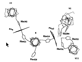
Figure 1.
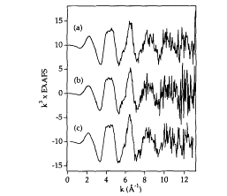
Figure 2.
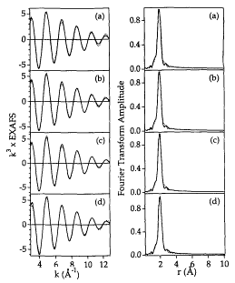
Figure 3.
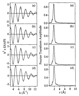
Figure 4.
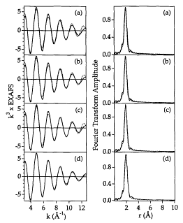
Figure 5.
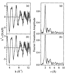
Figure 6.
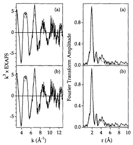
Figure 7.
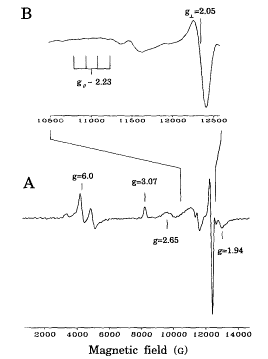
Figure 8.
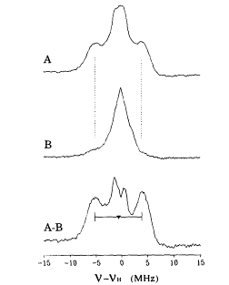
Figure 9.
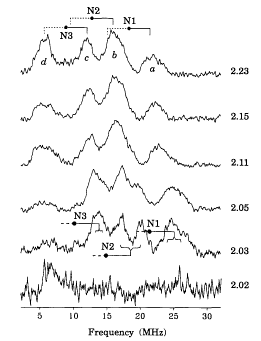
Figure 10.
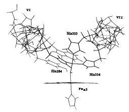
Figure 11.
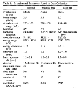
Table 1.
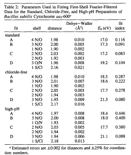
Table 2.
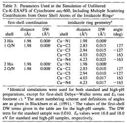
Table 3.
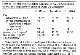
Table 4.
This page was last built with Frontier on a Macintosh on Thu, Jul 17, 1997 at 5:51:22 PM. Thanks for checking it out! John Boswell