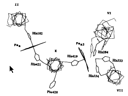
Figure 1. Model of the arrangement of the Fea , Fea3 , and CuB centers in the heme-copper respiratory oxidase superfamily. 2 Transmembranous helices II, VI,VII, and X, with their associated metal centers, are viewed from the periplasmic side (outside) of the membrane, perpendicular to the membrane plane. The metal ligand histidines are highlighted. In addition, the well-conserved residues Phe420 in helix X and Trp280 and Tyr288 in helix VI are shown. The through-bond pathway connecting Fea with Fea3 is highlighted, and the shortest distance between the heme edges is about 11 Å.
This page was last built with Frontier on a Macintosh on Thu, Jul 17, 1997 at 5:51:35 PM. Thanks for checking it out! John Boswell